
Bouveret syndrome:A case report
2019-04-22 09:01:42FeiWangZhiQiangDuYiLanChenTianMingChenYueWangXiangRongZhou
World Journal of Clinical Cases 2019年23期
Fei Wang, Zhi-Qiang Du, Yi-Lan Chen, Tian-Ming Chen, Yue Wang, Xiang-Rong Zhou
Fei Wang, Zhi-Qiang Du, Yi-Lan Chen, Tian-Ming Chen, Yue Wang, Xiang-Rong Zhou, Department of Gastroenterology, People’s Hospital of Jianyang City, Jianyang 641400, Sichuan Province,China
Abstract
Key words: Bouveret syndrome; Gallstones; Intestinal obstruction; Case report
INTRODUCTION
Bouveret syndrome is a rare clinical disease.Only 315 cases have been reported in the literature between 1967 and 2016.The disease is easy to get misdiagnosed and has high mortality if the diagnosis is delayed.Tools that have proven useful for diagnosing this disease include upper gastrointestinal endoscopy, computed tomography (CT) of the abdomen, and magnetic resonance cholangiopancreatography (MRCP).Here, we describe a case of Bouveret syndrome,which was initially misdiagnosed as gastric calculus based on endoscopy but was later correctly diagnosed with the help of CT.The patient was successfully treated by surgery after failed endoscopic therapy.
CASE PRESENTATION
Chief complaints
A 61-year-old woman presented with recurrent epigastric pain for more than 6 mo.Her abdominal pain had worsened during a period of 4 days and was associated with vomiting and abdominal distention.She denied having a fever with chills or jaundice.
History of past illness
Her previous history included hypertension and diabetes.
Personal and family history
The family history was unremarkable.
Physical examination
On clinical examination, her abdomen was soft with mild epigastric tenderness and positive succussion splash.
Laboratory examinations
Blood examination showed a white blood cell count of 13.1 × 10⁹/L with neutrophilia(75.7%), total bilirubin level of 28.7 mmol/L, direct bilirubin levels of 7.8 mmol/L,alanine aminotransferase level of 14 U/L, and aspartate aminotransferase level of 23 U/L.
Imaging examinations
On gastroscopy, a large amount of food residue was present in the gastric fundus and body, and multiple stones and ulcers were found in the pyloric region and first part of the duodenum, causing gastric outlet obstruction (Figure 1).A provisional diagnosis of gastrolithiasis was made.Endoscopic extraction of the stones using a snare was attempted but was unsuccessful.
Abdominal ultrasonography showed a 6.4 cm × 3.6 cm heteroechoic lesion in the region of the junction between the pylorus and duodenum with a 4.9 cm stone located within.The gallbladder was shrunken and located close to the heteroechoic lesion.Abdominal CT showed chronic cholecystitis with a cholecystoduodenal fistula and the presence of an approximately 3.0 cm × 2.4 cm stone in the descending part of the duodenum (Figure 2).There was partial intrahepatic bile duct dilatation with pneumobilia.
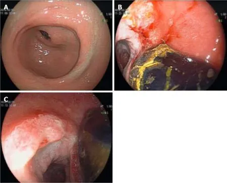
Figure 1 Endoscopic findings.
FINAL DIAGNOSIS
Based on the above findings, Bouveret syndrome was suspected.
TREATMENT
Repeat gastroscopy was performed, which showed a duodenal bulb stone with incarceration and a fistulous opening in the duodenal bulb (Figure 3).Due to the failure of multiple endoscopic stone extraction attempts, the patient had a hepatobiliary surgery consultation.Considering the patient’s long course of disease,CT findings of cholecystoduodenal fistula, and possibility of severe adhesions, open cholecystectomy with duodenal stone extraction and fistula repair were planned over laparoscopic treatment.
Intraoperatively, dense adhesions were present between the gallbladder and surrounding omentum tissue.The gallbladder was shrunken and thickened up to 1 cm with the presence of a cholecystoduodenal fistula of 2 cm.Calot’s triangle was frozen, making the dissection difficult.Upon opening the duodenum, an oval calculus about 4 cm x 6 cm in size was found in the descending duodenal segment.Cholecystectomy with duodenal stone extraction and fistula repair were performed.
OUTCOME AND FOLLOW-UP
The operation lasted 5 h and the intraoperative blood loss was 300 mL.The postoperative course was uneventful with a postoperative hospital stay of 7 d.At the last follow-up 6 mo after surgery, the patient was symptom-free.
DISCUSSION
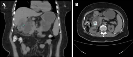
Figure 2 Computed tomography.
Gallstones can migrate into the intestinal tractviathe cholecysto- or choledochalduodenal fistula and block the proximal duodenum or pylorus, causing gastric outlet obstruction, a condition known as Bouveret syndrome[1].It is a rare complication of cholelithiasis.In the past 50 years from 1967 to 2016, only about 315 cases have been reported in the literature.The mortality rate of Bouveret syndrome is 12%–30% as it frequently occurs in elderly patients and is associated with many complications[2].Fistula formation occurs due to pressure necrosis of the gallbladder wall and the adjacent organ (duodenum in this case) densely adhered to it.Other contributory factors include chronic inflammatory changes in the gallbladder wall and impaction of the stone in the gallbladder neck[3].In the present case, the 6-mo history of epigastric pain was probably caused by calculous cholecystitis, and the calculi broke into the duodenal bulb after a long time.
Bouveret syndrome should be suspected in elderly patients with a previous history of chronic calculous cholecystitis, and sudden abdominal pain that is mainly in the right hypochondriac or epigastric region and is accompanied by nausea, vomiting,fever, and chills.Physical examination may reveal epigastric, subxiphoid, or right hypochondriac tenderness.Blood examination may show leukocytosis, deranged liver function tests, and elevation of inflammatory markers such as C-reactive protein.However, in rare cases it may show air-fluid levels suggestive of gastrointestinal obstruction, a high-density shadow in the right upper abdomen, and pneumobilia[4].
X-ray examination plays a limited role in diagnosing this disease.However,abdominal ultrasound is useful in determining the gallbladder wall thickening,gallbladder atrophy, and the presence or absence of gallstones.Moreover, in some cases, it can detect discontinuity in the gallbladder wall and the presence of gas in the gallbladder suggestive of internal fistula.Abdominal CT is an important tool for diagnosing this disease.The radiological triad of Rigler (small bowel obstruction,pneumobilia, and ectopic gallstone) is specific for its diagnosis[5,6].The so-called“ectopic gallstone” is the stone shadow on the right side at the level of T12–L1 vertebrae.MRCP, especially contrast-enhanced MRCP, has high diagnostic value as it can detect the presence of fistula, as well as the size and location of the stone and its relationship with the fistula[7].Upper gastrointestinal endoscopy is both diagnostic and therapeutic for this disease, as it can visualize the site and cause of upper gastrointestinal obstruction including the incarcerated stones.In about 31% of cases,the internal opening of the cholecystointestinal or choledochointestinal fistula can be detected[8].Sometimes, the huge stone blocks the fistulous opening and also limits the endoscopic vision, leading to misdiagnosis as was seen in the current case.
Various treatments for Bouveret syndrome have been described in the literature,and can be broadly divided into non-surgical and surgical treatments.Non-surgical treatment has a low success rate about 9%[9].However, with advances in endoscopic techniques, the success rate has greatly improved in the recent years.Common methods used to break the large stone into pieces include endoscopic lithotripsy,extracorporeal shock wave lithotripsy, electro-hydraulic lithotripsy, and endoscopic laser lithotripsy.The latter two technologies are simple, safe, and effective, with few complications, and are currently the most commonly used methods[10].However,some scholars believe that endoscopic lithotomy increases the risk of esophageal injury, digestive tract perforation, and gastrointestinal bleeding.A study showed that 42% of patients failed to achieve stone removal by this technique[11].Surgery is the mainstay of treatment and includes gastric or intestinal enterolithotomy with gallbladder duodenal fistula repair and cholecystectomy.It can be performed in a one or two-step method depending upon the patient’s age, physical condition, and disease characteristics.If the patient is in good general health, has a short history of symptoms, no major water, electrolyte imbalances, and no obvious inflammatory reaction at the site of choledocho- or cholecysto-intestinal fistula, one-stage operation can be considered[12].The advantages of this method are that it prevents the need for a second operation and prevents the occurrence of retrograde biliary tract infection,gallbladder cancer, and recurrence of gallstone ileus.However, because the majority of patients with Bouveret syndrome are elderly, with a long history of systemic diseases, the incidence of postoperative complications and mortality of one-step surgery is relatively high.In such patients with higher risk of surgical complications,a two-step method is beneficial in which only the gastric or intestinal stones are removed in the first surgery to relieve the obstruction.After the patient’s physical condition improves, fistula repair and cholecystectomy can be performed in the second stage.Postoperative complications and mortality of the two-step method are significantly lower than those of the one-step method.Moreover, in some cases, a second surgery is not required as the diseased gallbladder is already shrunken and lacks stones due to the fistula.In addition, some scholars believe that in most patients,the biliary fistula closes spontaneously after the intestinal calculi are removed, making it unnecessary to repair the fistula.In such cases, biliary symptoms are often relieved or even disappear in the absence of stones, despite leaving the gallbladderin situ[13].In recent years, with the extensive application of laparoscopy, laparoscopic surgery for Bouveret syndrome has become a safe and effective alternative to open surgery[14].
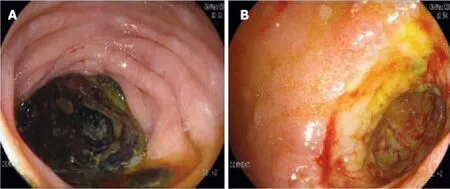
Figure 3 Repeat gastroscopy.
CONCLUSION
Bouveret syndrome is a rare clinical disease associated with high mortality, which requires a high index of clinical suspicion for timely diagnosis.CT, MRCP, and gastroscopy are the most useful tools for diagnosing this syndrome.Treatment should be individualized according to the patient’s age, physical condition, and disease characteristics.In view of the advantages of endoscopic lithotripsy or minimally invasive lithotripsy or lithotomy, it is recommended that endoscopic therapy be tried first; surgery can be performed if it fails[8].For patients with multiple comorbidities and poor general condition, the two-step surgical method can significantly reduce mortality[15].
 World Journal of Clinical Cases2019年23期
- World Journal of Clinical Cases的其它文章
- Pure squamous cell carcinoma of the gallbladder locally invading the liver and abdominal cavity:A case report and review of the literature
- Management of massive fistula bleeding after endoscopic ultrasound-guided pancreatic pseudocyst drainage using hemostatic forceps:A case report
- Fatal complications in a patient with severe multi-space infections in the oral and maxillofacial head and neck regions:A case report
- Left armpit subcutaneous metastasis of gastric cancer:A case report
- Rigid esophagoscopy combined with angle endoscopy for treatment of superior mediastinal foreign bodies penetrating into the esophagus caused by neck trauma:A case report
- Mixed serous-neuroendocrine neoplasm of the pancreas:A case report and review of the literature