
Effect of Nitric Oxide on Esophageal Cancer Cell Line TE-1
2013-11-18 13:33:30GuoguiSunWanningHuJunZhangChenglinLiandCongrongYang
Chinese Medical Sciences Journal 2013年1期
Guo-gui Sun ,Wan-ning Hu *,Jun Zhang ,Cheng-lin Li ,and Cong-rong Yang
1Department of Chemoradiotherapy,Tangshan People’s Hospital Affiliated to Hebei United University,Tangshan 063001,China
2Department of Radiology,Fourth Hospital of Hebei Medical University,Shijiazhuang 050011,China
ESOPHAGEAL cancer is a malignancy of digestive system with poor prognosis.Despite continuous improvement of conventional cancer treatment,the efficacy of early and long-term treatment for esophageal cancer remains unsatisfactory.Exploring and developing new therapies for esophageal cancer has become a critical clinical topic.In recent years,nitric oxide(NO) has been found to be a new radiosensitizing substance that can enhance the killing effect of radiation on tumor cells by reducing the intracellular antioxidant capacity.1A research indicates that NO is related to the radiosensitivity of oral squamous cell carcinoma and other malignancies.2However,its relationship with radiosensitivity of esophageal cancer has rarely been reported.This study employed a variety of techniques,including methyl thiazolyl tetrazolium (MTT) assay,flow cytometry (FCM),reverse transcription polymerase chain reaction (RT-PCR),and Western blot to assess the radiosensitizing effects of NO on esophageal cancer cells TE-1,particularly on cell proliferation,life cycle,and apoptosis.The potential mechanism underlying the sensitizing effects of NO combined with radiation were explored,thus providing valuable data for clinical treatment for esophageal cancer.
MATERIALS AND METHODS
Materials
Human esophageal cancer cell line TE-1 was provided by the Research Center of the Fourth Clinical Medical College,Hebei Medical University.MnSOD rabbit anti-human monoclonal antibody was purchased from Epitomics (Burlingame,Ca,USA),lyceraldehyde-3-phosphate dehydrogenase (GAPDH) mouse anti-human monoclonal antibody from Santa Cruz (Santa Cruz,CA,USA),IRDye 700 labeled goat anti-rabbit IgG antibodiy from LICOR bioscience(Lincoln,NE,USA),Trizol RNA extraction kit from Solarbio(Beijing,China),Superscript III reverse transcription kit purchased from Invitrogen (Grand Island,NY,USA).TaqDNA polymerase,OligodT,dNTPs,reverse transciptase,and RNases inhibitor were purchased from Fermentas(Glen Burnie,MD,USA).NO donor (sodium nitroprusside,SNP) was purchased from Beyotime Institute of Biotechnology (Beyotime,Guangzhou,China).
Cell growth inhibition and radiosensitivity measurement
The TE-1 cells were irradiated with linear accelerator with a 6 MV X-ray beam (irritation area,20 cm × 20 cm;source target distance,100 cm;gantry angle,180°),with 5 cm solid water placed under the 96-well culture plate.The TE-1 cells characterized by adherent monolayer growth were cultured in the high-glucose medium (DMEM) supplemented with 10% fetal calf serum at 37°C in 5% CO2.After 48-hour incubation,the cells were harvested,counted,and inoculated unto a 96-well culture plate (200 μL/well,5×103cells/μL).After 24-hour incubation,the cells were intervened with different concentrations of NO (0.1,0.5,1,1.5,2,and 4 mmol/L),different levels of radiation (1,2,4,6,and 8 Gy),or 1 mmol/L NO+2 Gy radiation for 12,24,48,and 72 hours,respectively,and then incubated for another 48 hours.Four hours before the incubation was ended,20 μL MTT (5 μg/mL) was added to each well.Four hours later,150 μL of 10% dimethylsulfoxide was added to each well,followed by oscillation for 10 minutes.
The optical density (OD) of intervened cells in each well was assayed with an automatic microplate reader.The detection wavelength and reference wavelength were 570 nm and 620 nm,respectively.Tumor cell inhibition rate (%)was calculated as∶[1-(experimentalODvalue/controlODvalue)]×100%.Tumor cell survival rate (%) was calculated as∶experimentalODvalue/controlODvalue ×100%.The cell curves were fitted to a multi-target single-hit model.In the formula,survival fraction (SF)=1-(1-e–D/D0)N,Dq=D0×lnN.SF=(Dvalue of experimental group/Dvalue of control group) × 100%,Dstands for the radiation dose,D0for the mean lethal radiation dose,Dqfor the quasithreshold dose (shoulder width of the survival),andNfor the extrapolated value.Sensitization enhancement ratio(SER)=D0of the single radiation group/D0of the NO+radiation group.
FCM detecting TE-1 cell apoptosis and cell cycle
Annexin V-PI double staining was used to detect the changes in TE-1 cell apoptosis after 48-hour treatment with 1 mmol/L NO,2 Gy radiation,or 1 mmol/L NO+2 Gy radiation.The cells were digested by trypsin,harvested,washed twice with 4°C pre-cooled phosphate buffered saline,and re-suspended with 1 mL buffer solution (final concentration of cells,1×106/mL).Cell suspension of 100 μL was added into a 5 mL FCM tube,to which 5 μL of Annexin V-FITC and 10 μL of 20 μg/mL propidium iodide were added.The mixture was incubated in the dark at room temperature for 15 minutes before FCM assay.
To detect the cell cycle,propidium iodide dye containing RNase was added to the cell suspension produced by re-suspending the digested,harvested,and washed cells in 1 mL buffer solution (final concentration of cells,1×106/mL).The mixture was incubated in the dark at room temperature for 30 minutes before assayed with FCM to determine the fluorescence intensity.
RT-PCR of manganese superoxide dismutase (Mn-SOD) mRNA
MnSOD is an important antioxidant enzyme in human cells.It can be used to transform superoxide radicalinto H2O2.Under the combined action of glutathione peroxides and catalase,H2O2is decomposed into water and oxygen,making the cells non-toxic.3Lower MnSOD expression in the cells may lead to the increase in reactive oxygen species (ROS) in mitochondria.High levels of ROS can cause the damage of DNA,proteins and lipids,and also increase the instability of the genome,which will cause tumor occurrence and help in normal cell transformation.4
For reverse transcription,total RNA was extracted from esophageal cancer TE-1 cells using Trizol and reverse transcription reagents following the manufacturer’s instructions.The extracted RNA was treated with 1 mmol/L NO,2-Gy radiation,or 1 mmol/L NO+2 Gy radiation for 48 hours,followed by reverse transcription to cDNA as the template for PCR amplification.The sequence of the upstream primer of MnSOD gene was 5'-AAGGTCGGAGTCAACGGATT-3',and that of the downstream primer of MnSOD gene was 5'-GCTCCTGGAAGATGGTGAT-3' (length of the primer fragment,158 bp).GAPDH was used as the internal reference (upstream primer,5'-CCGACC TGCCCTACGACTA-3';downstream primer,5'-CTGGGCTGTAACATC TCCCTT-3';length of primer fragment,226 bp).The PCR conditions were as follows∶pre-denaturation at 95°C for 3 minutes,35 cycles of denaturation at 95°C for 30 seconds,annealing at 56°C for 30 seconds,and elongation at 72°C.PCR products were qualified,and semiquantitative products were purified with 1.5% agarose gel electrophoresis.The OD value of purified products was analyzed with a gel imaging system and Multi-Gauge version 3.1.
Western blot of MnSOD protein
Western blot was conducted to determine the protein expression level of MnSOD in esophageal cancer TE-1 cells treated with 1 mmol/L NO,2 Gy radiation,or 1 mmol/L NO+2 Gy radiation for 48 hours.Four types of protein were collected from the RIPA protein lysate of harvested cells.The protein products were loaded to sample wells (50 μg/well)and purified using sodium dodecyl sulfate-polyacrylamide gel electrophoresis.The purified samples were placed in a stable ice bath,and then electrically transferred to a nitrocellulose membrane.The membrane was enclosed with 5%skim milk for 2 hours and incubated with the first antibody at 4°C overnight (MnSOD 1∶500,GAPDH 1∶500).The goat anti-rabbit IgG and infrared fluorescent secondary antibodies (1∶20 000) were added for continuous incubation at room temperature for 1 hour.The protein bands of Western blot were scanned and quantitatively analyzed with a twocolor infrared laser imaging system.
Statistical analysis
The experimental results were analyzed using SPSS 13.0.The data were expressed as means±SD.Single-factor analysis of variance (Ftest) was performed for intergroup comparison of cell apoptosis,cell cycle,cell growth inhibition rate,MnSOD mRNA expression,and MnSOD protein expression.P<0.05 was considered statistically significant.
RESULTS
Inhibitory effect of NO on TE-1 cell proliferation
MTT assay demonstrated that the cell inhibition rate was elevated along with the increase of NO concentration.Significant increase was observed in the inhibition rate of treatment with >1.0 mmol/L SNP.Among the cells treated with ≥0.5 mmol/L NO,significant differences were observed among the treatments for 12,24,48,and 72 hours with the same NO concentrations (P<0.05,Table 1).
Inhibitory effect of radiation on TE-1 cell proliferation
MTT assay demonstrated that the cell inhibition rate was elevated along with the increasing radiation dose (1,2,4,6,and 8 Gy) and duration (12,24,48,and 72 hours).The TE-1 cell inhibition rate was significantly increased when treated with radiation ≥2 Gy,and significant differences were observed among the treatment of 12,24,48 and 72 hours (P<0.05,Table 2).
Radiosensitivity of TE-1 cells
The cell survival curves fitted to a multi-target single-hit model showed that theD0,Dq,andNof TE-1 cells treated with 1,2,4,and 6 Gy radiation for 48 hours were 26.40,0.78,and 20.64,respectively.By comparison,those of the TE-1 cells treated in combination with 1.0 mmol/L NO for 48 hours were lower (9.84,0.50,and 4.91,respectively).Associated SER was therefore calculated as 2.68(Fig.1).
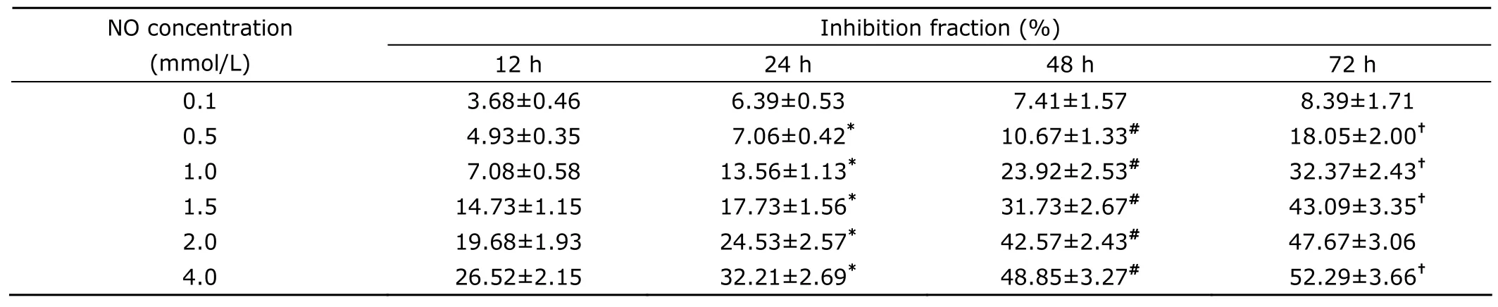
Table 1.The effect of NO on cell proliferation of human esophageal cancer cell line TE-1 detected by MTT§
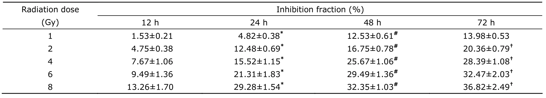
Table 2.The effect of radiation on TE-1 cell proliferation detected by MTT§
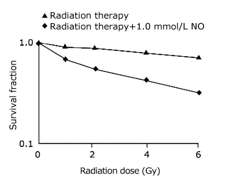
Figure 1.Radiobiological parameters of the cell survival curves fitted to a multi-target single-hit model.
Combined effect of NO and radiation on TE-1 cell apoptosis
As shown in the scatterplot of bivariate FCM,treatment of NO,radiation,or NO+radiation for 48 hours resulted in early cell apoptosis in the lower right quadrant (Fig.2).The proportion of apoptotic cells under the action of NO+radiation (10.7%±0.7%) was significantly higher than that under the action of NO (6.2%±0.4%,P<0.05) or radiation(10.7%±0.7%,P<0.05) alone.
Combined effect of NO and radiation on TE-1 cell cycle
After treatment of NO,radiation,or NO+radiation for 48 hours,the ratio of TE-1 cells in G2+M phase under the action of NO+radiation was higher than those in the cells treated with NO or radiation alone,while the ratios of TE-1cells in the G0+G1and S phases were relatively lower.Statistically significant differences were observed in the ratio of TE-1 cells at different cycle stages between NO +radiation-treated cells and NO-treated cells,also between NO+radiation-treated cells and radiation-treated cells(P<0.05,Table 3).
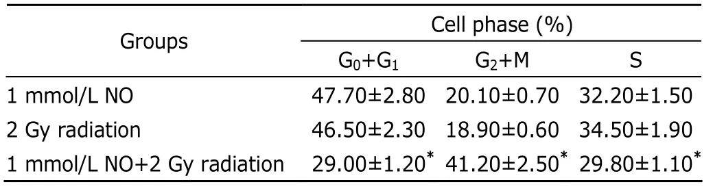
Table 3.TE-1 cell cycle detected by FCM§
MnSOD mRNA expression and protein expression
RT-PCR showed that the MnSOD mRNA expression level in TE-1 cells treated with NO+radiation for 48 hours was 0.12±0.03,significantly lower than that in the cell treated with NO (0.21±0.02,P<0.05) or radiation alone (0.20±0.01,P<0.05) (Fig.3).
Western blot result showed that the MnSOD protein expression level in TE-1 cells treated with NO+radiation for 48 hours was 0.13±0.1,significantly lower than that in the cells treated with NO (0.23±0.2,P<0.05) or radiation alone (0.24±0.3,P<0.05) (Fig.4).
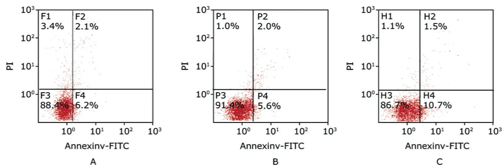
Figure 2.The effects of NO and radiation on TE-1 cell apoptosis assayed by FCM.
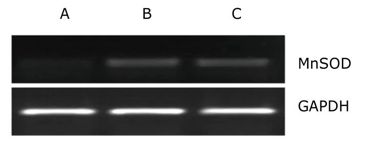
Figure 3.The effects of NO and radiation on manganese sup-eroxide dismutase (MnSOD) mRNA expression of TE-1 cells.
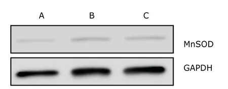
Figure 4.The effects of NO and radiation on the MnSOD pritein expression of TE-1 cells.
DISCUSSION
Esophageal cancer is a common human gastrointestinal malignancy with a 5-year survival rate of only about 20%.5The main cause of the failure of radiotherapy is the uncontrolled or recurrent tumors in the local radiation field,which suggests the presence of radiation-resistant tumor cells.Therefore, one of the research focuses in this field is to develop an effective radiosensitizer for improving the radiosensitivity of esophageal cancer cells, further enhancing the efficacy of radiotherapy.
NO is generated from L-arginine by the catalysis of nitric oxide synthase.It features high activity of free radicals and is involved in various biological processes.A study has shown that high NO level can induce the apoptosis of nerve cells, macrophages, and tumor cells.6However, the underlying functional mechanism remains unclear.Results of this study showed that treatment with different levels of NO and/or different doses of radiation affected esophageal cancer cell line TE-1 by inhibiting cell proliferation, promoting cell apoptosis, and increasing the SER.With >0.5 mmol/L NO, the inhibitory effect on TE-1 cell proliferation was significantly enhanced, reflecting the concentration- and dose-effect relationship, consistent with previous findings regarding the proliferation inhibition and apoptosis promotion effects of NO on gastric cancer cell lines AGC and BGC-823.6,7In addition, the SER of V79 cells and SCK cells treated with NO donor reached 2.6 and 2.8, respectively,1,8indicating that NO significantly affected the radiosensitivity of tumor cells.FCM assay detecting the cell cycle of esophageal cancer TE-1 cells showed that the ratio of cells in the G2+M phase after 1.0 mmol/L NO +2 Gy radiation treatment was higher than that after 1.0 mmol/L NO or 2 Gy radiation treatment alone, whereas the ratio of TE-1 cells in the G0+G1and S phase was decreased after 1.0 mmol/L NO+2 Gy radiation treatment.Similarly, a previous study in China showed that the cycle of SGC-7901 cells changed after treatment with sodium nitroprusside,with an increased proportion of cells in the G0/G1phases and a decreased proportion of cells in the S and G2/M phases.6Komatsu et al9found that NO increased the radiosensitivity of mouse fibrosarcoma cells (FSa-II) with doxorubicin-induced MnSOD cDNA.RT-PCR and Western blot in this study confirmed that the expression levels of MnSOD mRNA and protein were reduced after treated with NO, radiation, or NO + radiation, and this effect was mostly significant with NO + radiation treatment.In the study by Wang et al,10the sensitivity of human osteosarcoma cell line to doxorubicin at different levels of MnSOD showed a bidirectional regulation.That is, high MnSOD expression level decreased the sensitivity by decreasing cell apoptosis while low MnSOD expression level increased the sensitivity by enhancing cell apoptosis.It was suggested that H2O2produced by different MnSOD expressions possibly reacted with other reactive oxygen species to generate different chemicals.
In conclusion, this study demonstrated that NO significantly affected esophageal cancer cells by inhibiting cell proliferation, promoting cell apoptosis, and enhancing radiosensitivity.These effects may be related to the prevention of cell cycle and inhibition of the expression of MnSOD mRNA and protein.
1.Mitchell JB, Cook JA, Krishna MC, et al.Radiation sensitisation by nitric oxide releasing agents.Br J Cancer Suppl 1996; 27∶S181-4.
2.Dong XY, Zhao SF, Zhu FD, et al.Apoptosis induces by exogenous nitric oxide in Tca8113 cells.Zhejiang Da Xue Xue Bao Yi Xue Ban 2006; 35∶ 50-4.
3.Sun GG, Wang YD, Chen LQ, et al.Novel cancer suppressor gene for esophageal cancer∶ manganese superoxide dismutase.Dis Esophagus 2011; 24∶346-53.
4.Orrenius S,Gogvadze V, Zhivotovsky B. Mitochondrial oxidative stress∶ implications for cell death. Annu Rev Pharmacol Toxicol 2007; 47∶ 143-83.
5.Schuchert MJ, Luketich JD, Landreneau RJ.Management of esophageal cancer.Curr Probl Surg 2010; 47∶845-946.
6.Sang JR,Cheng YC,Shao GB,et al.Effect of nitric oxide on the proliferation of AGC gastric cancer cells.Chin J Cancer 2010;29∶158-62.
7.Lin W,Song JD,Lin JY.Nitric oxide effect of proliferation of stomach cancer cell line BGC-823.Chin J Cancer 2001;20∶1043-8.
8.Janssens MY,Verovski VN,Van den Berge DL,et al.Radiosensiti-zation of hypoxic tumour cells by S-nitroso-Nacetylpenicillamine implicates a bioreductive mechanism of nitric oxide generation.Br J Cancer 1999;79∶1085-9.
9.Komatsu M,Kuroda M,Wang Y,et al.Manganese superoxide dismutase overexpression changes plating efficiency bidirectionally according to change in redox for a human osteosarcoma cell line,SaOS2.Int J Oncol 2005;26∶853-62.
10.Wang Y,Kuroda M,Gao XS,et al.Hydrogen peroxide overload increases adriamycin-induced apoptosis of SaOS(2)FM,a manganese superoxide dismutase-overexpressing human osteosarcoma cell line.Int J Oncol 2005;26∶1291-300.
 Chinese Medical Sciences Journal2013年1期
- Chinese Medical Sciences Journal的其它文章
- Clinical Study on Suspension Pancreatic-Duct-Jejunum End-to-Side Continuous Suture Anastomosis in Pancreaticoduodenectomy
- Awareness of Cornea Donation of Registered Tissue Donors in Nanjing△
- Breast Milk Lead and Cadmium Levels in Suburban Areas of Nanjing,China
- Possible Role of Mast Cells and Neuropeptides in the Recovery Process of Dextran Sulfate Sodium-induced Colitis in Rats△
- Effect of Phenylephrine on Alveolar Fluid Clearance in Ventilator-induced Lung Injury△
- Arthroscopic Debridement and Synovium Resection for Inflammatory Hip Arthritis