
Late infection after peri-orbital autologous micro-fat graft:a case presentation and literature review
2024-03-20 06:33:44FarzadPakdelHassanAsadigandomaniMelikaArabBafraniZohrehNozarianZohreAbedinifarNiloofarPirmarzdashtiBehzadJafariZaynabSiami
Farzad Pakdel, Hassan Asadigandomani, Melika Arab Bafrani, Zohreh Nozarian,Zohre Abedinifar, Niloofar Pirmarzdashti, Behzad Jafari, Zaynab Siami
1Department of Oculofacial Plastic Surgery, Farabi Hospital,School of Medicine, Tehran University of Medical Sciences,Tehran 1336616351, Iran
2Craniomaxillofacial Research Center, Tehran University of Medical Sciences, Tehran 1336616351, Iran
3Department of Ophthalmology, School of Medicine, Tehran University of Medical Sciences, Tehran 1336616351, Iran
4Department of Pathology, Farabi Hospital, School of Medicine, Tehran University of Medical Sciences, Tehran 1336616351, Iran
5Department of Microbiology, Farabi Hospital, School of Medicine, Tehran University of Medical Sciences, Tehran 1336616351, Iran
6Pediatric Cell and Gene Therapy Research Center, Children’s Medical Center, School of Medicine, Tehran University of Medical Sciences, Tehran 1416753955, Iran
Dear Editor,
Currently, autologous micro-fat graft (AFG) to periorbital area is widely used to address cosmetic and functional issues[1].Infections after AFG are rare.The overall prevalence of infection after AFG has been reported to be 0.043%[2].Infection typically occurs during 2wk after fat injection[3].To the best of our knowledge, late infection AFG has been rarely addressed in the literature[4].There are few reports on late infections after micro-fat graft with atypical microorganisms,including mycobacteria and fungal species[5-6].Delayed infection has been reported after non-facial cosmetic AFG procedures such as breast augmentation[7].Early infections after AFG are caused by typical microorganisms includingStaphylococcus epidermidisandStaphylococcus aureus.In this study we report a rare occurrence of clinical features,challenges in diagnosis and successful management of a patient case with late consecutive bilateral infection following periorbital cosmetic micro-fat injection in an otherwise healthy patient.The study adhered to the tenets of the Declaration of Helsinki.Written consent was obtained for publishing patient’s identifiable images and photographs.
Case ReportA 48-year-old female was referred to senior oculo-facial plastic surgeon (Pakdel F) for right lower eyelid swelling and redness in March 2, 2022.She had underwent autologous fat injection for correction of infra-orbital hollow about three months before our visit.Fat injection had been performed in an office.Harvest site was abdomen.
She had no remarkable past medical history.Initial examinations showed an otherwise healthy middle-aged woman with swelling and hard violaceous erythema in the right lower eyelid-check with mild tenderness (Figure 1A).Systemic examination showed one submandibular lymph node non-tender enlargement.Initial systemic examination,complete blood count, erythrocyte sedimentation rate (ESR)and C-reactive protein (CRP) were within normal limits.Computed tomography (CT) scan was compatible with preseptal cellulitis (Figure 2A).
She had history of bilateral AFG in December 1, 2021.Forty days after injection, in January 10, 2022, she experienced swelling with mild erythema and mild pain in the right lower eyelid and received oral cephalexin for ten days and then coamoxiclav for one month.Upon no improvement, she had received oral prednisolone and also intralesional triamcinolone by other physicians.She reported a transient decrease in erythema and swelling after triamcinolone injection followed by even more severe erythema, swelling and pain.Thereafter,she experienced progressive worsening despite different treatment plans with oral co-amoxiclav, and then one-week intravenous cephazolin as an out-patient treatment, and continued oral prednisolone 50 mg per day.Based on the clinical manifestations, late and indolent course of the infection and our previous experiences we considered the possibility of atypical mycobacterial infection and also biofilm associated infection of the microfat graft.The patient was admitted to the hospital and received intravenous 500 mg vancomycin every eight hours, intravenous piperacillintazobactam (Tazocin) 4.5 g every eight hours and oral azithromycin 500 mg every day.No remarkable change was observed within three days after admission and intravenous antibiotics.Magnetic resonance imaging supported micro abscesses in the lower eyelid-cheek area (Figure 2B).We performed a conservative debridement of the necrotic and micro abscess loci and removed infected nidus and also took tissue biopsy for bacteriology and pathology studies through transconjunctival approach by senior author (Pakdel F) in March 5, 2022.Result of the culture showedStaphylococcal spp.and was negative for acid-fast bacilli.Multinucleated giant cells were found in the hematoxylin and eosin (H&E) staining of the tissue specimens.No distinct necrotizing granulomatous reaction was found.Ziehl-Neelsen staining and polymerase chain reaction (PCR) study on the tissue specimen for atypical mycobacteria were negative.She showed gradual improvement within three days after surgery.The patient was discharged with oral cephalexin 500 mg every 6h and oral azithromycin 500 mg per day.Erythema and induration of the right side was reduced significantly and slowly.The inflammation settled down within four weeks.
She suddenly developed erythema, swelling and induration of the contralateral left lower eyelid-cheek area in May 9, 2022(Figure 1B).The patient declared that she did not take her medication for last one week.She was admitted to hospital and received intravenous vancomycin and intravenous tazocin.Spiral orbital CT scan showed well-defined periorbital soft tissue swelling and hypodense loci compatible with micro abscess corresponded with whitish lower eyelid skin spot on erythematous context.She underwent a meticulous debridement of the necrotic material through transconjunctival approach by the senior author (Pakdel F).
Culture of the debrided specimen showed coagulase-negativeStaphylococci spp.Histology exam with H&E staining showed mature congested adipose tissue with mild chronic inflammation.She showed a remarkable resolution of periorbital erythema and swelling within next two weeks.She continued oral azithromycin 500 mg for three months.In six months follow up, peri-orbital swelling, erythema and pain were resolved.There was a mild skin irregularity and mild bilateral post-inflammatory hyperpigmentation over lower eyelid-cheek (Figure 1C).Both the patient and her primary physician expressed their satisfaction.The patients expressed that she was happy enough to do any more intervention for correction of he mild irregularity of the lower eyelid-cheek junction.
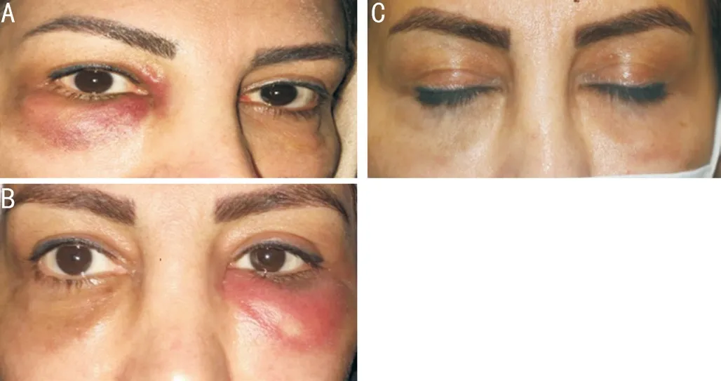
Figure 1 Face photograph A: Right lower eyelid-cheek erythema and swelling denoting a pre-septal cellulitis.Small deep grayish loci compatible with micro-abscesses.B: Late lower eyelid -cheek erythema and swelling denoting a pre-septal cellulitis.Small deep grayish loci compatible with micro-abscesses.C: Resolved erythema and swelling on the both sides after surgery and medical treatment.Post inflammatory hyperpigmentation and mild residual irregularity.

Figure 2 Imaging studies A: CT scan.Axial view.Softtissue swelling and fat stranding in lower eyelid-cheek area with small hypo dense areas.B: Magnetic resonance imaging, T1 weighted with gadolinium.Axial view.Diffuse soft tissue enhancement involving cutaneous,fascia and muscular layers.Small hypointense areas were seen consistent with micro-abscess.
DISCUSSION
Infection after AFG is unusual.Furthermore, late infection is extremely rare.A systematic review on 5577 patients that received AGF showed infection in 0.06% and 1.04%of patients with cosmetic and reconstructive procedures,respectively[2].In a systematic review on breast augmentation with autologous fat graft, infection was reported in 7.03% of all complications[8].Conde-Greenet al[9]reported infection in 8.33% of all complications after gluteal fat augmentation.Thus, it seems that the rate of infection varies on the location of augmentation with breast and gluteal areas carrying higher risk than face.
Postoperative infections are commonly due toStaphylococcalspecies and often resolve with a short course of oral antibiotics[10].Staphylococcushas been the causative agent for late infections after knee, breast, and cardiac prosthesis.They account substantially for foreign body-related infections.Coagulase negativeStaphylococcus(CoNS) have a strong capacity to form biofilm.It can, at least partly, explain the late occurrence of infection and resistance to antibiotic treatment[10].The route of infection in our case, as in other similar graft associated infections, was considered as intra-operative contamination.This could be inoculation through skin, adnexa or contaminated instruments.This explains the necessity of sterile setup for fat harvest and injection by cannula.Culture of the biopsied tissue and debrided material in our case showedStaphylococcalinfection as the main or part of the causative organisms.Biofilm in the contaminated microfat graft explains a late and bilateral involvement in our patient.
There are reports that atypical mycobacteria includingMycobacteriumfortuitum andAspergillusfumigatus infections may also cause late infection after AFG[11].It often presents as multiple nodules or mass like lesion in the peri orbital area.Orbital cellulitis may occur subsequently.The mean time of clinical presentation is about 12wk, although some studies suggest a short interval, ranging from 18 to 24d[12].
Despite laboratory and bacteriology methods for diagnosis of atypical mycobacterial infections, still clinical manifestations and response to empirical treatment seem reliable[13].Noncaseating granuloma, multinucleated giant cells, necrosis,Ziehl-Neelsen staining method and fluorescent microscopy are all helpful for diagnosis of the causative agents.Positive culture on mycobacterial-specific growth media is the gold standard for diagnosis, but clinical manifestations of suspected cases of this infection is enough for initiation of treatment.Negative PCR cannot rule out mycobacterial infection, because a lot of confounding factors can be effective on the result of this test, such as lack of organisms on the surface of the lesion and a low number of microorganisms in the infected tissue[14].A case series of thirteen patients reported atypical mycobacterial infection after ophthalmic reconstructive surgeries[15].In one study 12 out of 13 patients were treated with antibiotic therapy but one patient was treated with combination of antibiotics and surgical drainage.A number of non-infectious entities may be associated with early onset inflammation after fat graft[16-22].Our patient showed clinically significant resolution of swelling and erythema after meticulous debridement and azithromycin.The patient had recurrent infection after 1mo of antibiotic therapy.Based on the above in delayed infections after periocular procedures,Staphylococcaland atypical mycobacteria should be considered.
In conclusion, late infection of AFG in the peri-orbital is a rare and complex entity.Timely, correct diagnosis and appropriate management need understanding of this possibility and appropriate surgical and antibiotic treatment.It is reasonable to suggest that fat grafting to be preferably performed under sterile conditions in a standard operating room.Physicians need to be cautious about local injection of corticosteroids in cases with fat graft late inflammation, before making sure about the absence of any indolent infectious process.
ACKNOWLEDGEMENTS
Conflicts of Interest:Pakdel F,None;Asadigandomani H,None;Arab Bafrani M,None;Nozarian Z,None;Abedinifar Z,None;Pirmarzdashti N,None;Jafari B,None;Siami Z,None.
 International Journal of Ophthalmology2024年3期
- International Journal of Ophthalmology的其它文章
- Stromal lenticule addition keratoplasty with corneal crosslinking for corneal ectasia secondary to FS-LASlK:a case series
- Clinical features and possible pathogenesis of multiple evanescent white dot syndrome with different retinal diseases and events: a narrative review
- Utility of real-time 3D visualization system in the early stage of phacoemulsification training
- Efficacy of scleral buckling for the treatment of rhegmatogenous retinal detachment using a novel foldable capsular buckle
- Effect of navigation endoscopy combined with threedimensional printing technology in the treatment of orbital blowout fractures
- Outcomes and variables that impact pneumatic retinopexies