
Magnetic resonance imaging features of intrahepatic extramedullary hematopoiesis: Three case reports
2022-12-19 08:06:52MaLuoJiaWenChenChuanMiaoXie
World Journal of Clinical Cases 2022年19期
Ma Luo, Jia-Wen Chen, Chuan-Miao Xie
Abstract
Key Words: Liver; Extramedullary hematopoiesis; Signal intensity; Magnetic resonance imaging; Case report
lNTRODUCTlON
Extramedullary hematopoiesis seldom occurs within the liver alone[1]. In this rare condition, the lesion can manifest as a mass with no typical radiologic findings, making it difficult to diagnose and differentiate from other hypervascular neoplasms[2]. We present three cases of intrahepatic extramedullary hematopoiesis (IEMH) occurring solely in the liver. These lesions showed lower signal intensity on inphase images than on out-phase images. In addition, the first case was unique in that the lesion showed changes in magnetic resonance imaging (MRI) signal intensity with size enlargement between two rounds of imaging examination. These manifestations have never been reported before.
CASE PRESENTATlON
Chief complaints
Case 1: A 50-year-old woman without any discomfort was admitted to our hospital due to an intrahepatic mass with interval growth.
Case 2: A 30-year-old female with a five-month history of Hodgkin’s lymphoma (nodular sclerosis) was referred to hospital.
Case 3: A 52-year-old male was admitted to our hospital due to the incidental discovery of hepatic nodules. He had no history of alcoholism.
History of present illness
Case 1: Negative.
Case 2: The patient denied alcoholism and had no other symptoms or discomfort.
Case 3: No symptoms or discomfort.
History of past illness
Negative.
Personal and family history
Case 1: The patient’s medical history included thyroid carcinoma and lung adenocarcinoma. She had undergone total thyroidectomy in November 2014 and lobectomy of the right lower lung lobe in October 2019, without radiotherapy or adjuvant chemotherapy in the subsequent follow-up.
Case 2: Prior to the patient’s initial treatment, no focal liver lesion was detected by ultrasound or positron emission tomography (PET)/computed tomography (CT). She had undergone 5 cycles of chemotherapy consisting of Brentuximab Vedotin + Adriamycin, Vinblastine (Dacarbazine).
Case 3: The patient had no previous medical and family history.
Physical examination
Case 1: Physical examination was normal.
Case 2: Physical examination was normal. The liver and spleen were impalpable.Case 3: Physical examination was negative.
Laboratory examinations
Case 1: Laboratory tests, including blood, liver function, and serum tumor marker tests (alpha fetoprotein, carbohydrate antigen 19-9, carbohydrate antigen 125, carcinoembryonic antigen, and protein induced by Vitamin K absence or antagonist-II), were all within the normal range. Hepatitis serologic markers such as hepatitis B surface antigen and hepatitis C virus antibodies were negative, with no history of alcoholism.
Case 2: Platelet, white, and red blood cell counts were normal. The serum tumor markers and hepatitis serologic markers were negative.
Case 3: Laboratory tests revealed that liver function, serum hepatic tumor markers, and hepatitis serologic markers were all normal.
Imaging examinations
Case 1: In September 2019, pre-operative ultrasound showed a hyperechoic lesion measuring 24 mm × 23 mm in segment VIII. The lesion was diagnosed as possible hemangioma.
In January 2020, during a routine examination, the lesion showed an increase in size on abdominal ultrasound. An abdominal CT scan (Discovery 750 HD, GE Healthcare, Milwaukee, WI, United States) was performed. The lesion appeared heterogeneously hypodense on unenhanced CT (40 HU), and 35 mm × 32 mm × 20 mm in size. The lesion was moderately hyperdense in the arterial phase (81 HU) and markedly hyperdense in the portal venous phase (106 HU), and showed persistent enhancement in the 5-min delayed phase (110 HU) (Figure 1A-D). The following day, liver MRI was performed for further characterization and assessment of the lesion (Trio 3.0 T, Siemens, Erlangen, Germany). The lesion was hypointense relative to surrounding liver tissue on T1 weighted images (WI) with and without fat saturation (FS), and was heterogeneously hyperintense on T2WI or T2WI-FS with clear demarcation. On dynamic contrast enhancement series, the lesion showed considerable enhancement in the arterial phase, remaining hyperintense relative to surrounding liver tissue in both the portal venous and delayed phases (6 min) following the injection of 0.1 mmol/kg of Gd-DTPA (Magnevist, Bayer Healthcare, Berlin, Germany) (Figure 1E-K). The initial diagnosis included atypical angioleiomyolipoma, adenoma, and single metastasis. The patient declined to undergo surgery and opted for observation.
In June 2020, she underwent another abdominal MRI examination. The lesion had enlarged to 50 mm × 46 mm × 43 mm, and it was heterogeneously hypointense on T1WI-FS with scattered foci of hyperintense areas. The signal intensity was significantly lower on the in-phase image than on the outphase image. The lesion was isointense or mildly hypointense relative to liver tissue on diffusion weighted images (DWI), T2WI, and T2WI-FS. The pattern of enhancement was overt in the arterial phase and remained hyperintense on the delayed phase, without vessel encasement or invasion (Figure 1L-R).
Case 2: In April 2020, abdominal ultrasound was performed during a routine examination and a slightly hyperechoic lesion was found in segment VII. The patient then underwent an MRI scan (Discovery 750W, GE Healthcare, Milwaukee, WI, United States). A 10 mm × 8 mm sized, slightly hyperintense and hypointense lesion was identified on T2WI-FS and T1WI-FS, respectively (Figure 2A and B). The signal intensity was lower on the in-phase image than that on the out-phase image (Figure 2C and D). The signal intensity was lost on susceptibility weighted imaging (SWI) (Figure 2E). After administration of Gd-DTPA, the lesion showed poor enhancement in the arterial phase and enhanced gradually and persistently up to 6 min (Figure 2F-H). The initial diagnosis was lymphoma infiltration.
Case 3: MRI (Trio 3.0 T, Siemens, Erlangen, Germany) scans showed a lesion located in the transitional area (34 mm × 27 mm in size) between segment V and VIII, with homogeneously low and high signal intensity on T1WI-FS and T2WI-FS, respectively. The signal intensity on the in-phase image was lower than that on the out-phase image (Figure 3A-E). After administration of Gd-EOB-DTPA, the lesion showed intense enhancement in the arterial phase, with no persistent enhancement in the portal venous and transitional phases, and was hypointense in the hepatobiliary phase (Figure 3F-H). The possible diagnosis at that time included hepatoma, adenoma, and angiomyolipoma.

Figure 1 An intrahepatic mass (arrow) in segment Vlll was found in a 50-year-old female. A: On unenhanced computed tomography, the lesion was heterogeneously hypodense, with hyperdense foci in the central area; B: The lesion was heterogeneously hyperdense in the arterial phase; C and D: Progressive enhancement in the portal venous (C) and delayed phases (D); E-K: On magnetic resonance imaging, the lesion was heterogeneously hyperintense on T2 weighted image (WI), T2WI-fat saturation (FS) (E and F) and diffusion weighted imaging (DWI) (G), isointense on the apparent diffusion coefficient map (H), hypointense on T1WI-FS (I), avid enhancement in the arterial phase (J) and persistent enhancement in the delayed phase (K); L-R: Corresponding follow-up images five months later showed that the lesion size had increased. Signal drop was seen on the in-phase (L) compared with the out-phase (M) image. The lesion was heterogeneously hypointense on T2WI-FS (N) and DWI (O), homogeneously hypointense on T1WI-FS (P), and showed hypervascular enhancement with delayed enhancement in the arterial (Q) and delayed phase (R), respectively. The surgical pathologic diagnosis was intrahepatic extramedullary hematopoiesis.
FlNAL DlAGNOSlS
Case 1: Macroscopically, a mass with reddish appearance without necrosis or hemorrhage was seen in the resected specimen. Microscopically, megakaryocytes, and erythroid cells were scattered within the hepatocyte cords (Figure 4). Immunohistochemical staining including CD3, CD20, CD61, CD235, and MPO was performed, and the cells were positive for these markers. These findings were consistent with EMH.
Case 2: Percutaneous fine-needle aspiration biopsy was performed under ultrasound guidance, and cytology showed EMH.
Case 3: The final histopathological diagnosis was EMH (Figure 5).

Figure 2 A 30-year-old female diagnosed with intrahepatic extramedullary hematopoiesis was confirmed by biopsy. A and B: The lesion (arrow) located in the subcapsular of segment VI/VII was slightly hyperintense on T2 weighted image (WI)-fat saturation (FS) (A) and slightly hypointense on T1WI-FS (B); C-E: The lesion showed a lower signal intensity on the in-phase (C) than on the out-phase (D) image, and signal loss on susceptibility weighted imaging (E); F-H: In dynamic series, the lesion was mildly enhanced in the arterial phase (F), with areas of progressive and prolonged enhancement in the portal venous (G) and delayed phases (H).

Figure 3 A 52-year-old male diagnosed with intrahepatic extramedullary hematopoiesis was confirmed by resection. A-D: The lesion located in segment V/VIII (arrow) showed lower signal intensity on the in-phase (A) than on the out-phase (B) image, as opposed to another lesion (the same patient) in segment IV (arrowhead, the surgical pathologic diagnosis was angioleiomyolipoma), which showed higher signal intensity on the in-phase (C) than on the out-phase (D) image; E-H: The lesion (arrow) showed high signal intensity on T2 weighted image-fat saturation (E), with intense enhancement in the arterial phase (F), and was relatively hypointense in the transitional phase (G) and hepatobiliary phase (H).
TREATMENT
Case 1: The patient underwent hepatic segmentectomy.
Case 2: After another 4 cycles of chemotherapy, liver MRI (August 2020) showed similar findings, without any increase in the size of the lesion. As the diagnosis was uncertain and the lesion was small, we decided to obtain histopathologic confirmation.
Case 3: As the possibility of malignancy could not be completely excluded, the patient underwent hepatic segmentectomy, although he did not have any predisposing factors for hepatoma.
OUTCOME AND FOLLOW-UP
Case 1: The patient has no recurrent lesions or evidence of new lesions.
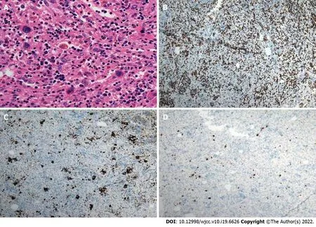
Figure 4 lntrahepatic extramedullary hematopoiesis in the same patient shown in figure 1. A: On the photomicrograph (hematoxylin and eosin staining; × 200), megakaryocytes and erythroid cells were scattered within the surgical specimen; B-D: Immunohistochemical staining using CD235 (B), CD61 (C) and MPO (D) markers (× 40) revealed that the cells were positive (brown color) for these markers, respectively.
Case 2: The patient has no recurrent lesions or evidence of new lesions.
Case 3: The patient has no signs of recurrence.
DlSCUSSlON
IEMH is a rare, benign condition of the liver. The radiological literature on this disease is exclusively case reports. All of these cases were misdiagnosed, and IEMH was not considered in the preoperative imaging diagnosis and differentiation. The most common imaging modalities used were CT and ultrasound[3-7]. There is a paucity of literature on the role of MRI, which might be due to the limitations of imaging technology at that time. Moreover, the quality of the images provided in those studies was low. Thus, the radiologic features seemed to be non-specific, and it may have been difficult to make a correct diagnosis preoperatively.
From the pathological aspect, EMH is characterized by numerous hematopoietic cells. Granulocytes, megakaryocytes, and erythrocytes show a mixed distributed within the lesion, in which adipocytes and fibrous tissue can also be seen. Thus, EMH is positive for MPO, CD61, and CD235. Due to the large number of erythrocytes scattered within EMH, CD235 staining is strong and diffusely positive. The presence of iron in the erythrocytes and different amounts of adipose tissue can potentially affect the radiologic appearance.
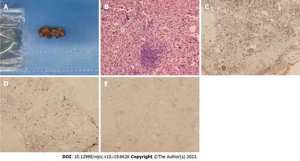
Figure 5 lntrahepatic extramedullary hematopoiesis in the same patient shown in figure 3. A: Photograph of the specimen showed the lobular and solid nature of the resected hepatic mass (segment V/VIII), without areas of necrosis and hemorrhage; B: On the photomicrograph (hematoxylin and eosin staining; × 200), granulocytes, megakaryocytes, adipocytes and erythrocytes were distributed within the surgical specimen; C-E: Immunohistochemical staining using CD235 (C), CD61 (D) and MPO (E) markers (× 40) revealed that the cells were positive (brown color) for these markers, respectively.
In recent years, with the development of MRI, there have been sporadic reports regarding the published MRI findings of IEMH. Belayet al[8] provided the first description of T2*WI in their case, considering that this technique might have a potential role in MRI diagnosis. The lesion was hypointense on T2*WI, similar to the signal intensity of the hepatic background on this gradient recalled echo (GRE) sequence, indicating that the lesion had as much iron deposition as the liver parenchyma in the setting of secondary hemochromatosis owing to repeated blood transfusions. Leeet al[2] reported the superparamagnetic iron oxide (SPIO)-enhanced MRI in their case, while Zhang and Zhu[9] applied the chemical shift images in their report. In our three cases, lesions on in-phase images exhibited lower signal intensity than on out-phase images. In Case 2, the lesion was hypointense in SWI. In Case 1, there were signal changes across two time intervals on T1WI, T2WI, and DWI, which shifted from hyperintensity to hypointensity, and without restricted diffusion in two sets of apparent diffusion coefficient (ADC) maps. We speculate that these manifestations and signal changes may be mainly ascribed to iron deposition, depending on iron evolution in the lesion, instead of the increase in cell density or change in intralesional content such as mucin, necrosis, fibrosis, or even calcification. Iron could impact the magnetic field intensity and homogeneity; thus, the signal on DWI could be diverse due to iron content and evolution in IEMH, but not due to diffusion restriction on the ADC map. However, there are limited data regarding EMH on DWI. Rascheet al[10] observed that EMH in the spleen could impact the DWI signal. The GRE sequence was unequivocally sensitive to the presence of small amounts of iron. It should also be noted that, when the echo time was longer, the signal indicating iron deposition in the lesion grew less intense[11]. Such lesions often showed lower signal intensity on SWI and in-phase images relative to the out-phase images.
Some cases report using scintigraphy to diagnose IEMH[3,12,13], and the lesions showed Tc-99m uptake. In addition, PET was applied in the diagnosis of EMH located in the paraspinal region, peritoneum and lung[1,13,14]. High uptake values were observed. However, there are no reports on the PET characteristics of IEMH. We speculate that its property on18F-FDG might be from mild to intense activity, which may be related to the different stages of the disease. In the initial stage, the synthesis and proliferation of hematopoietic cells were active, thus presenting as hypermetabolism, whereas in the static stage, IEMH could show low uptake.
There are discrepancies regarding the radiologic characteristics in different studies (Table 1). IEMH was described as a fat-containing lesion. For example, in reports published by Guptaet al[15], Navarroet al[6] and Cao and Wang[16], multiple lesions showed fat density. However, the case by Zhang and Zhu[9] showed a solitary lesion without any fat content, as indicated by the lack of fat signal alteration. With regard to enhancement pattern, the lesion presented as a hypervascular mass with heterogeneous enhancement in the report by Wonget al[17] and homogeneous avid enhancement in the report by Zhang and Zhu[9], while mild enhancement was reported by Tammet al[12]. Elsayeset al[18] considered that an active lesion exhibited iso or hyperintensity on T1WI and T2WI, and was enhanced after injection of contrast material, while older lesions could be hypointense on T1WI and T2WI, and might show no enhancement. Nevertheless, Kumaret al[19] provided a case where the lesion was isointense on T1WI and T2WI, but showed almost no enhancement. Belayet al[8] showed a lesion with low signal intensity on both T1WI and T2WI with marked enhancement. Some authors have indicated that iron deposition and fat infiltration might refer to an old stage in the disease course[20], while other authors thought that iron and fat content were also detectable in the active stage[21]. MRI was found to reflect different stages of hematopoiesis, depending on iron signal evolution, fat content, degree of fibrous organization, and vascular enrichment in the lesion. In this way, the difference in hematopoietic materials, fibrous tissue and fat content could explain the diverse radiologic description and the lack of exclusive imaging patterns.

Table 1 Clinical and magnetic resonance imaging features of 9 cases with intrahepatic extramedullary hematopoiesis
Although most of the patients with IEMH had hematological disease, a few cases had no evidence of this underlying condition (as shown in the Case 1 and Case 3). For example, small cell lung cancer and Noonan syndrome were reported in two cases[4,15], and the cause of IEMH in another two cases remains unknown[22,23].
Two factors differentiate these three cases from those in other reports. First, no hepatomegaly or splenomegaly was found in these patients. Second, a change in MRI signal was observed at two different points in time in Case 1 and it presented more radiologic characteristics over the course of the disease. Interestingly, all our reported lesions were either in segment VII or VIII. IEMH in other segments have been reported in other cases. More cases should be documented in the future.
The differential diagnosis includes benign, primary, and secondary liver malignant lesions. IEMH may mimic these lesions leading to troublesome diagnosis. In the “fat deposition” stage, the characteristic signal intensity on the in-phase image is higher than that on the out-phase image. The differential diagnosis in cirrhotic liver includes fatty metamorphosis in hepatocellular carcinoma (HCC), while that in non-cirrhotic liver includes benign lesions such as focal fatty infiltration (without mass effect), adenoma (hepatocyte nuclear factor 1a-mutated subtype) and lipoma (no enhancement). Angiomyolipoma should be also taken into consideration. In the “iron deposition” stage, the characteristic signal intensity on the in-phase image is lower than that on the out-phase image. The differential diagnosis (intratumoral bleeding) includes benign lesions such as adenoma (inflammatory subtype) and hemangioma. Malignant lesions include hemorrhagic HCC and metastasis. When IEMH demonstrates strong and persistent enhancement, focal nodular hyperplasia, adenoma and hypervascular metastasis need to be considered. An appropriate clinical setting and the application of Gd-EOB-DTPA or SPIO are helpful in the diagnosis. When IEMH shows mild enhancement or avid enhancement with “washout”, atypical metastasis, HCC, or even fibrolamellar carcinoma in young patients should be considered in the differential list. Lymphoma is homogenous isointense with moderate enhancement. Fat and bleeding content is seldom seen in lymphoma.
CONCLUSlON
IEMH has a variable radiologic appearance and is easily misdiagnosed. Given its rarity and the lack of pathognomonic imaging findings, awareness of its presentations might help radiologists establish the diagnosis.
ACKNOWLEDGEMENTS
The authors thank Chao Zhang, MD, PhD, and Jing-Ping Yun, MD, PhD, for their contributions to the pathological figure and analysis.
FOOTNOTES
Author contributions:Luo M and Chen JW collected the data; Luo M, Chen JW and Xie CM analyzed the data; Luo M wrote the original draft; Xie CM reviewed and edited the manuscript; all authors have read and approved the final manuscript.
lnformed consent statement:Informed written consent was obtained from the patient for publication of this report and any accompanying images.
Conflict-of-interest statement:All the authors report no relevant conflicts of interest for this article.
CARE Checklist (2016) statement:The authors have read the CARE Checklist (2016), and the manuscript was prepared and revised according to the CARE Checklist (2016).
Open-Access:This article is an open-access article that was selected by an in-house editor and fully peer-reviewed by external reviewers. It is distributed in accordance with the Creative Commons Attribution NonCommercial (CC BYNC 4.0) license, which permits others to distribute, remix, adapt, build upon this work non-commercially, and license their derivative works on different terms, provided the original work is properly cited and the use is noncommercial. See: https://creativecommons.org/Licenses/by-nc/4.0/
Country/Territory of origin:China
ORClD number:Ma Luo 0000-0003-4581-3454; Jia-Wen Chen 0000-0002-9435-2108; Chuan-Miao Xie 0000-0001-8533-623X.
S-Editor:Fan JR
L-Editor:Webster JR
P-Editor:Fan JR
 World Journal of Clinical Cases2022年19期
- World Journal of Clinical Cases的其它文章
- Hem-o-lok clip migration to the common bile duct after laparoscopic common bile duct exploration: A case report
- Preliminary evidence in treatment of eosinophilic gastroenteritis in children: A case series
- Identification of risk factors for surgical site infection after type II and type III tibial pilon fracture surgery
- Sustained dialysis with misplaced peritoneal dialysis catheter outside peritoneum: A case report
- Delayed-onset endophthalmitis associated with Achromobacter species developed in acute form several months after cataract surgery: Three case reports
- Diagnostic accuracy of ≥ 16-slice spiral computed tomography for local staging of colon cancer: A systematic review and meta-analysis