
Lumbar disc sequestration mimicking a tumor: Report of four cases and a literature review
2022-06-22 08:37:08ShengTangLiTaoZhangXueWenShiHuaLiuChengWeiYangPingZhenSongKaiLi
World Journal of Clinical Cases 2022年9期
lNTRODUCTlON
Lumbar disc herniation is a common condition in orthopedics defined as the displacement of disc material beyond its anatomical space,and the formation of an isolated disc when the herniated disc is detached from the parent disc.Disc sequestration refers to the migration of herniated disc fragments into the epidural space;most disc fragments move in a lateral,cephalic,or caudal direction due to the anatomical structure of the anterior epidural space,and can be easily misdiagnosed as spinal tumors[1,2].In rare cases,fragments may migrate dorsally into the posterior epidural space or be located intradurally.The lumbar spine is the most commonly affected region,but the cervical and thoracic spine can also be affected.Here,we report four cases of disc sequestration mimicking a spinal tumor and review the literature to provide insight into discriminating between a herniated disc and spinal tumor.All four cases underwent surgery,and the postoperative pathology showed intervertebral disc tissue.All four cases had an uneventful postoperative course and recovered completely within 3 mo[3].
CASE PRESENTATlON
Chief complaints
A 71-year-old man was admitted for low back pain with left lower extremity radiating pain for 1 year,with exacerbation over the past 2 wk.
A 74-year-old man was admitted for pain in both knees and movement limitation for 3 years,with exacerbation over the past 3 mo.
She cast one more lingering, half-fainting glance at the prince, and then threw herself from the ship into the sea, and thought her body was dissolving into foam
A 53-year-old man was admitted for numbness and weakness of the lumbar spine and right lower extremity for 2 wk.
We have described four cases of intervertebral disc herniation.The herniated intervertebral discs were all in the epidural space.In two cases,they migrated to the posterior space and did not have characteristic imaging findings on contrast-enhanced MRI.Our preliminary diagnosis of the four cases was spinal tumor.The free intervertebral disc should be considered when differentially diagnosing spinal lesions.For patients who cannot be diagnosed before surgery,surgical resection of the lesion and pathological examination can aid diagnosis.
History of present illness
The patient had low back pain with left lower extremity radiating pain for 1 year.After intermittent acupuncture treatment,the symptoms were slightly relieved,but the symptoms were exacerbated by physical work performed 2 wk ago.There was no numbness or weakness in either lower extremity,and defecation and urinary functions were normal.
The pathogenesis of lumbar disc herniation may be related to adhesions that form between the ventral dura mater and posterior longitudinal ligament.Repeated minor traumas or previous surgery can exacerbate the adhesions.The anterior epidural space is limited to the posterior longitudinal ligament and sagittal midline diaphragm.It spans the space between the vertebral body and posterior longitudinal ligament,preventing protruding disc fragments from crossing the midline.The lateral membrane is attached to the posterior longitudinal ligament and extends laterally to embed into the lateral wall of the spinal canal.The lateral membrane and posterior longitudinal ligament limit backward movement of free disc fragments[1,5].Due to the anatomical structure of the anterior epidural space,most cases involve movement of intervertebral disc fragments in the lateral,cranial,or caudal direction[1,4,6-10].In rare cases,fragments move back into the posterior epidural space or are located in the dura mater[2,3,11-17].The lumbar spine is the most commonly affected area,but the intervertebral discs of cervical and thoracic vertebrae may be displaced[4,8,12].
Suddenly, everything grew quiet. My sister began to clear the table. My brother was scraping the last of the egg from his plate. And then that ancient family ritual that had filled so many Sunday mornings came to an end. My father announced: Let s go read the paper, Hon.
The patient had numbness and weakness of the lower back and right lower extremity for 2 wk.The sites of numbness of the right lower extremity were mainly the right hip,back of the thigh,and back of the lower leg,accompanied by an obvious limitation of movement.Walking 50 m required intermittent squatting,which relieved the symptoms.Physical therapy such as massage had a poor effect.Defecation and urinary functions were normal.
There was no obvious trigger for right lower extremity pain in this patient.There was obvious pain in the anterolateral thigh and knee joint of the right lower extremity,but this was not accompanied by lumbar pain or limited movement of either lower extremity.The patient had not undergone any treatment.The symptoms became more severe 1 wk ago,so a painkiller was administered at a local hospital.The symptoms were not significantly relieved,so he was transferred to our hospital.
History of past illness
None of the four patients had a previous history of diseases.
Personal and family history
None of the patients had any relevant personal or family history.
On previous trips we had seen all the famous monuments and tourist sights. The guidebooks claimed that locals were rude and indifferent to visitors but there had to be more to the people of Paris than that. This time we wanted to find the real Parisians.
Physical examination
Flexion and extension movements of the lumbar spine were slightly restricted,and there was mild tenderness over the L3-4,L4-5,and L5-S1 interspinous spaces.The muscle tone of both lower limbs was normal,the strength of the left gluteus maximus and iliopsoas muscles was grade IV,and the strength of the remaining muscles was normal.In the leg raising test,it was positive(50°)on the left lower limb.Knee and ankle reflexes were reduced bilaterally,but there were no pathological reflexes.
It was taken to the palace and tied at the foot of the Lady Jamila s raised seat, but she ordered a longer cord to be brought so that it might be able to jump up beside her
The patient was discharged on postoperative day 6 with fully recovered neurological function,and the preoperative pain symptoms had disappeared.He returned to normal life after the 3-mo followup.
Limping gait was present,lumbar flexion and extension were significantly limited,and the L4-5 and L5-S1 spinous processes and interspinous space were tender.Muscle tension in the lower limbs was normal.Muscle strength of the right lower limb was grade IV.The skin sensation of the right hip,posterolateral thigh,and lateral leg was decreased,while muscle strength and skin sensation of the left lower limb were normal.The bilateral straight leg raising test was normal.The left knee and ankle reflexes were weak,but were normal on the right side.There were no pathological reflexes.
Limping gait was present,and lumbar flexion and extension were limited,but muscle tension of both lower limbs was normal.Except for the iliopsoas muscle(grade III strength),the other muscles of the right lower limb were grade IV,and the muscle strength of the left lower limb was normal.The leg raising test was positive(60°).The right knee reflex was weak,and the bilateral Babinski sign was positive.
Laboratory examinations
All parameters in all four cases,including but not limited to complete blood count,renal and liver function tests,prostate-specific antigen,and other specific tumor markers,were within the normal ranges.
Imaging examinations
After admission,magnetic resonance imaging(MRI)showed an abnormal shadow in the spinal canal of the L4 vertebral segment and compression of the dural sac.T1-weighted images showed high signal intensity,while T2-weighted images showed low signal intensity,with heterogeneous enhancement on contrast-enhanced MRI(Figure 1A-1E).

After admission,an abnormal shadow on the right side of the L4-5 segment spinal canal was detected on MRI,compressing the cauda equina nerve and right nerve root.T1- and T2-weighted images showed low signal intensity.A “marginal capsule” and separation from the lesion were significantly enhanced on contrast-enhanced MRI(Figure 2A-2E).
The girl with him was quite cute, dressed in a white sailor s uniform, but her refreshing14 charm quickly faded from memory as I turned and encountered more students costumed to celebrate the various evil spirits representative of this night
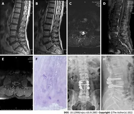
After admission,an abnormal shadow was observed in the right intervertebral foramen of the L3 vertebral canal on MRI,and the adjacent dural sac was compressed.The T1-weighted image showed moderate signal intensity,while the T2-weighted image showed high signal intensity.There was no obvious enhancement on contrast-enhanced MRI(Figure 4A-4E).

PRlMARY DlAGNOSlS
According to the results of the preoperative physical,laboratory,and imaging examinations,cases 1 and 2 were diagnosed with neurilemmoma,and cases 3 and 4 with spinal tumors.
I always dreamed of having a college education and now I m getting one. She told me. After class we walked to the student union building and shared a chocolate milk shake. We became instant friends. Every day for the next three months we would leave class together and talk nonstop. I was always mesmerized1 listening to this time machine as she shared her wisdom and experience with me.
FlNAL DlAGNOSlS
All four patients were diagnosed with lumbar disc herniation.
TREATMENT
The patient was discharged on postoperative day 5 with fully recovered neurological function,and the preoperative pain had disappeared.He returned to normal life after the 3-mo follow-up.
A laminectomy,discectomy,and internal fixation were performed(Figure 2G-2H).The focus was located in the right posterior epidural space of L4-5,which significantly compressed the dural sac.Postoperative pathology confirmed that the focus was intervertebral disc tissue(Figure 2F).
A laminectomy,discectomy,and internal fixation were performed(Figure 3G-3H).The focus was located in the right anterior L4-5 epidural space.Postoperative pathology confirmed that the focus was intervertebral disc tissue(Figure 3F).
A hemilaminectomy,discectomy,and internal fixation were performed(Figure 4G-4H).The focus was located in the right anterior epidural space,compressing the dural sac and right L3 nerve root.Postoperative pathology confirmed that the focus was intervertebral disc tissue(Figure 4F).

OUTCOME AND FOLLOW-UP
A laminectomy,discectomy,and internal fixation were performed(Figure 1G-1H).The focus was located in the left posterior epidural space,which compressed the dural sac and left L4 nerve root.Postoperative pathology confirmed that the focus was intervertebral disc tissue(Figure 1F).
After admission,an abnormal shadow was detected in the L4-5 segment spinal canal on MRI,and spinal canal stenosis was observed at the same level.High signal intensity was detected on the T2-weighted image,and peripheral enhancement was observed on contrast-enhanced MRI(Figure 3A-3E).
The patient was discharged on postoperative day 8 with fully recovered neurological function,and the preoperative pain symptoms had disappeared.He returned to normal life after the 3-mo followup.
Simon replied that the king of the country had insisted on giving him one of his daughters as a wife, but that he had refused the honour because he was too old and too frail37
The patient was discharged on postoperative day 8 with fully recovered neurological function,and the preoperative numbness symptoms had disappeared.He returned to normal life after the 3-mo follow-up.
Limping gait was present,and lumbar flexion and extension were slightly limited.Tenderness of the L4-5 spinous process and interspinous space was detected.Muscle strength and muscle tension were normal in both lower limbs.And the bilateral straight leg raising test was normal.The bilateral knee reflex was normal,but the bilateral ankle reflex was weak.There were no pathological reflexes.
DlSCUSSlON
Intervertebral disc herniation refers to displacement of the intervertebral disc outside its anatomical space.Disc sequestration is defined as the migration of protruding disc fragments into the epidural space and complete separation from the parent disc[1,2].Intervertebral disc herniation is closely related to degeneration of the intervertebral disc.When a herniated intervertebral disc prolapses into the epidural space,it expands rapidly because the intervertebral disc nucleus is rich in proteoglycans with strong hydrophilicity.The spinal cord and nerve root are easily compressed during the early stage of congestion,resulting in clinical symptoms[4].
The patient had pain in both knees with limited movement for 3 years,which was not significantly improved after intake of oral painkillers and intra-articular injection of sodium hyaluronate.The symptoms had been more severe for nearly 3 mo.Defecation and urinary functions were normal.
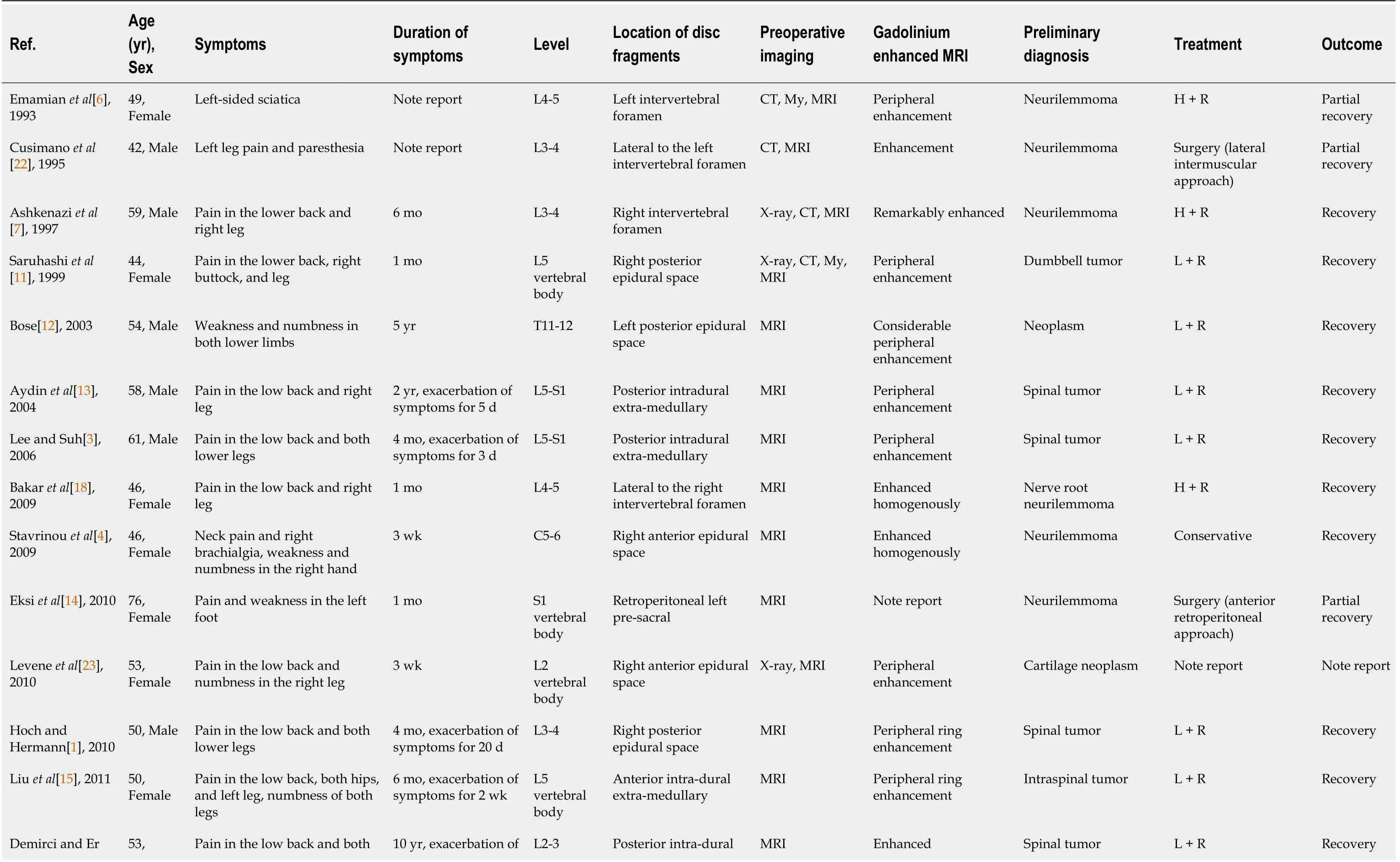
Because of the uncertainty of the anatomical location and atypical imaging features,the free disc fragments in the spinal canal are easy to misdiagnose as spinal tumors.We used the term “disc herniation,mimicking tumor” to search PubMed from 1990 to 2020,and retrieved 65 articles.After reading the full texts,we identified 23 highly relevant reports(24 cases)that gave detailed information on all patients(Table 1).This encouraged us to report our four cases.


Although MRI helps clinicians to accurately diagnose intraspinal soft tissue lesions,it lacks specificity[12].Therefore,it is necessary to distinguish prolapsed intervertebral disc tissue from epidural abscesses,dissolving epidural hematomas,synovial cysts,neurilemmomas,lipomas,and meningiomas.A free intervertebral disc in the spinal canal has low signal intensity on T1-weighted images and high signal intensity on T2-weighted images.Peripheral enhancement of non-enhanced intervertebral disc fragments is observed on contrast-enhanced MRI[8,17-19].An epidural abscess is often located in the posterior epidural space.Compared with the spinal cord,it produces low or moderate signal intensity on T1-weighted images and high signal intensity on T2-weighted images.Contrast-enhanced MRI shows homogenous or peripheral enhancement.Subdural hematomas have similar manifestations,while dissolving epidural hematomas have a circular appearance and enhancement.MRI signalintensity is similar to that of the cerebrospinal fluid,and there are “focal marks” on the spinal cord.If there is no enhancement on contrast-enhanced MRI,a synovial cyst is likely.Neurilemmomas are often located within the epidural space;they are isointense on T1-weighted images,hyperintense on T2-weighted images,and enhanced on contrast-enhanced MRI.More than half of lipomas occur in the dura mater.Lipomas display high signal intensity on T1-weighted images,low signal intensity on T2-weighted images,and low signal intensity on fat-suppressed images.Meningiomas are most common in the thoracic spine and are often located in the dura mater.They are isointense on T1- and T2-weighted images and enhanced on contrast-enhanced MRI[1,8,12].
He whined20 and sobbed21 out: Give, oh give me my beard again, and I will instruct you in all the magic art that surrounds this castle, and will help you to carry off the hidden treasure, which will make you rich and happy for ever
Case 1 showed high signal intensity on the T1-weighted image,low signal intensity on the T2-weighted image,and heterogeneous enhancement on contrast-enhanced MRI(Figure 1A-1E).Case 2 had low signal intensity on the T1-weighted image,low signal intensity on the T2-weighted image,and heterogeneous peripheral enhancement on contrast-enhanced MRI(Figure 2A-2E).These two cases were diagnosed as neurilemmomas before the operation.Case 3 displayed high signal intensity on the T2-weighted image and obvious peripheral enhancement on contrast-enhanced MRI(Figure 3A-3E).Case 4 exhibited moderate signal intensity on the T1-weighted image and high signal intensity on the T2-weighted image,without enhancement on contrast-enhanced MRI(Figure 4A-4E).These two cases were initially diagnosed as spinal tumors before the operation.Due to the nonspecific imaging findings,we were unable to diagnose these four patients by computed tomography,MRI,or enhanced MRI,so surgery was performed.The focus and adjacent intervertebral disc were resected,the intervertebral space was bone-grafted,and internal fixation was performed.The resected lesions were sent for pathological diagnosis.All four cases were pathologically diagnosed as herniated disc tissue(Figure 1F-4F).
Disc fragments usually show peripheral rim enhancement on contrast-enhanced MRI,which is related to the inflammatory reaction and new blood vessels around the intervertebral disc fragments.The degree of peripheral rim enhancement depends on the degree of angiogenesis[1,20,21].However,atypical enhancement or non-enhancement on enhanced MRI made diagnosis difficult in this study.
Imaging is not ideal for diagnosing soft tissue masses.Even when the imaging diagnosis seems clear,pathological diagnosis is still the gold standard[7].
We describe and analyze the detailed characteristics of tumor-like disc herniations evident on contrast-enhanced MRI,and we sought to distinguish them from other spinal diseases with which herniations are easily confused.However,after reviewing the literature,we cannot draw a clear conclusion.We speculate that when peripheral enhancement is evident on contrast-enhanced MRI,a herniated disc should be strongly suspected.In the future,we will seek to enhance the accuracy of MRI and contrast-enhanced MRI used to diagnose intervertebral disc herniation.
CONCLUSlON
A 75-year-old man was admitted for pain in the right lower extremity for 2 wk,with exacerbation over the last week.
FOOTNOTES
Li ST and Li SK provided the concept for the study and drafted the manuscript;Zhang T and Shi XW provided the images;Liu H,Yang CW,and Zhen P performed the operations,all authors have read and approved the content of the manuscript.
Chinese People’s Liberation Army Medical Technology Youth Training Program,No.20QNPY071.
So the Tsar and Vasilissa the Beautiful were married, and her father returned from the far-distant Tsardom, and he and the old woman lived always with her in the splendid Palace, in all joy and contentment. And as for the little wooden doll, she carried it about with her in her pocket all her life long.
When he was very nearly home he again thought of the cow that was with calf9, so he laid down the money, ran home, and asked his wife whether the cow had calved yet
Informed consent was obtained from the patients for publication of the case report.
It took a long time to find myself, and I had to live alone to do it. But I am not lonely. I am free for the first time in my life. I am Tandaleah, the Fire Goddess of the Volcano, spelled with two Ds and I m living happily ever after.
The authors declare no conflicts of interest for this article.
The authors have read the CARE Checklist(2016),according to which the manuscript was prepared and revised.
“It is gold! it is gold!” cried they, rushing forward, and seizing the horses. Then they struck the little jockeys, the coachman, and the footman dead, and pulled little Gerda out of the carriage.
This article is an open-access article that was selected by an in-house editor and fully peer-reviewed by external reviewers.It is distributed in accordance with the Creative Commons Attribution NonCommercial(CC BYNC 4.0)license,which permits others to distribute,remix,adapt,build upon this work non-commercially,and license their derivative works on different terms,provided the original work is properly cited and the use is noncommercial.See: http://creativecommons.org/Licenses/by-nc/4.0/
China
Sheng-Tang Li 0000-0002-6004-1934;Tao Zhang 0000-0003-2308-094X;Xue-Wen Shi 0000-0001-8360-3453;Hua Liu 0000-0003-0593-9001;Cheng-Wei Yang 0000-0001-6130-4874;Ping Zhen 0000-0003-3122-4042;Song-Kai Li 0000-0002-9200-2390.
Xing YX
My Days are really hectic7 and when I come home I don t pay a lot of attention to you. Sometimes I scream at you for not getting good enough grades in school and for your bedroom being a mess, but somehow tonight, I just wanted to sit here and well, just let you know that you do make a difference to me. Besides your mother, you are the most important person in my life. You re a great kid and I love you!
Wang TQ
Xing YX
 World Journal of Clinical Cases2022年9期
- World Journal of Clinical Cases的其它文章
- Malignant struma ovarii with papillary carcinoma combined with retroperitoneal lymph node metastasis:A case report
- Upper gastrointestinal bleeding from a Mallory-Weiss tear associated with transesophageal echocardiography during successful cardiopulmonary resuscitation:A case report
- lpsilateral hemifacial microsomia with dextrocardia and pulmonary hypoplasia:A case report
- Esophageal myoepithelial carcinoma:Four case reports
- Turner syndrome with primary myelofibrosis,cirrhosis and ovarian cystic mass:A case report
- Acute coronary artery stent thrombosis caused by a spasm:A case report