
Portal hypertension in a patient with biliary hamartomas:A case report
2020-05-13 07:34:48QianQianLiXiaoZhongGuoHongYuLiXingShunQi
World Journal of Clinical Cases 2020年9期
Qian-Qian Li, Xiao-Zhong Guo, Hong-Yu Li, Xing-Shun Qi
Qian-Qian Li, Xiao-Zhong Guo, Hong-Yu Li, Xing-Shun Qi, Department of Gastroenterology,General Hospital of Northern Theater Command, Shenyang 110840, Liaoning Province, China
Qian-Qian Li, Postgraduate College, Dalian Medical University, Dalian 116044, Liaoning Province, China
Abstract
Key words: Biliary hamartomas;Portal hypertension;Variceal bleeding;Computed tomography;Magnetic resonance imaging;Case report
INTRODUCTION
Biliary hamartomas (BH), also known as von Meyenburg complexes, are usually considered a benign disease caused by congenital bile duct malformation[1,2].They are clinically rare with a prevalence of 0.6% on biopsy[3].Microscopical images often show bile duct-like structures covered by a single layer of columnar epithelium.Dilated lumens contain bile and are surrounded by fibrous stroma[2,4].Except for liver biopsy,BH can often be detected by computed tomography (CT) and magnetic resonance imaging (MRI) images, which often appear as multiple irregularly shaped lesions with a diameter of about 10 mm[5].
Most patients with BH are usually asymptomatic.Some patients accidentally present with mild symptoms, such as abdominal pain, fever, or liver dysfunction[1,6,7].Herein, we report a patient with BH who presented with variceal bleeding and underwent endoscopic variceal therapy.
CASE PRESENTATION
Chief complaints
On September 20, 2018, a 40-year-old man presented with dark red colored bloody stool for one day.
History of present illness
The patient presented with dark red colored bloody stool for one day.He had been diagnosed with esophageal and gastric varices on endoscopy, and then underwent endoscopic variceal ligation and repeated gastric glue tissue adhesive injection for variceal bleeding at our department.
History of past illness
He had no history of hepatitis virus infection, alcohol abuse, drug-induced liver injury, or autoimmune liver disease.
Laboratory examinations
At admission, laboratory tests showed that hemoglobin was 55 g/L, red blood cell count was 1.90 × 10¹²/L (reference range:4.3-5.8 × 10¹²/L), hematocrit was 16.3%(reference range:40%-50%), white blood cell count was 3.0 × 10⁹/L (reference range:3.5-9.5 × 10⁹/L), platelet count was 21 × 109/L (reference range:125-350 × 109/L),prothrombin time was 16.8 s (reference range:11.5-14.5 s), and activated partial thromboplastin time was 36.7 s (reference range:28.0-40.0 s).Other biochemical indices showed no obvious abnormalities.He received intravenous infusion of proton pump inhibitors and vasoconstrictors and a transfusion of suspended red blood cells and fresh frozen plasma.
Imaging examinations
On September 21, 2018, the patient underwent upper gastrointestinal endoscopy,which showed mild esophageal varices, portal hypertensive gastropathy, and a removing tissue glue at the gastric fundus which was considered the major source of gastrointestinal bleeding.Thus, our endoscopist did not perform endoscopic variceal therapy on this patient.Contrast-enhanced CT scans showed multiple, rounded, low density areas on the liver, which were not significantly enhanced at the arterial and portal vein phases (Figure 1).MRI and magnetic resonance cholangiopancreatography(MRCP) were then performed.MRI showed rounded, irregular, low-signal-T1 and high-signal-T2 lesions diffusely distributed on the liver which were not significantly enhanced (Figure 2).MRCP showed that cystic high-signal lesions were diffusely distributed on the liver but were not communicated with the biliary system (Figure 3).The patient refused liver biopsy.
FINAL DIAGNOSIS
A diagnosis of BH was considered.
TREATMENT
He continued to receive intravenous infusion of proton pump inhibitors,vasoconstrictors and nutritional fluids.Following treatment, he was stable and discharged on oral propranolol.
OUTCOME AND FOLLOW-UP
On December 5, 2018, the patient was again admitted to our department due to melena for 5 h.Upper gastrointestinal endoscopy showed mild gastric varices and portal hypertensive gastropathy which was considered the major source of gastrointestinal bleeding.
On February 11, 2019, the patient underwent upper gastrointestinal endoscopy,which showed mild esophageal varices and several gastric varices with red color sign.He then underwent endoscopic glue tissue adhesive injection.
On July 4, 2019, the patient underwent upper gastrointestinal endoscopy.At this time, endoscopic variceal therapy was not necessary.He underwent MRI and MRCP again.No significant changes in liver lesions were seen when compared with the previous images (Figures 4 and 5).A diagnosis of BH was still considered.
DISCUSSION
Congenital malformations of the intrahepatic bile duct include many diseases, such as BH, Caroli disease, autosomal dominant polycystic kidney disease, and congenital hepatic fibrosis, which have malformations at different levels of the biliary tree.Of these, BH refers to malformations of small interlobular bile ducts[8,9].BH are characterized as multiple low-density lesions with an irregular outline on plain CT scans with a diameter of 10-15 mm.They are not usually enhanced[9].Generally, MRI is considered to be superior to CT in the diagnosis of BH[10,11].On MRI scans, BH lesions are characterized as low signal intensity on T1-weighted images and high signal intensity on T2-weighted images[12,13].In a few cases, the rim of lesions can be enhanced, which is considered to be the surrounding compressed liver parenchyma[14,15].BH should be distinguished from several other diseases.First,simple hepatic cysts, a benign disease, are characterized as round homogeneous lowdensity lesions with typically regular outlines without enhancement on CT scans.Hepatic cysts are larger in size than BH[10].Second, Caroli disease is a congenital malformation of larger intrahepatic bile ducts.Multiple low-density lesions can be seen on CT scans.The classical imaging presentation is the central dots of the cystic bile duct surrounding the branches of the veins.MRCP is usually a good diagnostic modality for Caroli disease, which can show that the cystic bile duct communicates with the biliary system[16,17].Third, polycystic liver disease is inherited and often associated with autosomal dominant polycystic kidney disease.Multiple cystic lesions are often observed in both liver and kidney[18,19].Due to a large number of cysts, the incidence of intracapsular hemorrhage is increased, and abnormal signals can be seen on the images[10].Positive family history is also one of the major diagnostic criteria[20].Fourth, cystic metastases are characterized as an enhancement of peripheral viable tissue around the lesion on contrast-enhanced CT and MRI scans[10].Fifth, abscesses are inflammatory lesions characterized as double target signs on CT, which consist of a hypodense pus area, a hyperdense ring of granulation tissue, and a hypodense zone of inflammatory edema[21].In addition, abscesses can be readily differentiated from BH by clinical symptoms, such as fever and abdominal pain[22].
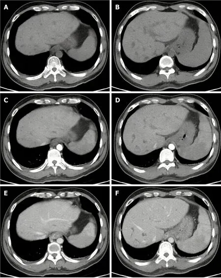
Figure 1 Computed tomography images of biliary hamartomas on September 21, 2018.
Due to the small diameter of lesions, BH may not lead to obvious clinical symptoms.Only a few studies have reported that BH might progress to cholangiocarcinoma[23].However, our patient had another rare but fatal portal hypertension-related complication, and presented with bleeding from gastroesophageal varices and hypersplenism.Of course, the etiology of portal hypertension in this case needs to be differentiated from liver cirrhosis which is often produced by viral hepatitis, alcohol abuse, or autoimmune factors.However, the patient refused to undergo liver biopsy.In the absence of liver histological analyses,we would like to emphasize that our case does not have any potential cause of chronic liver diseases nor abnormal liver function.Therefore, we believe that portal hypertension was mainly related to the presence of BH.Indeed, Yoshidaet al[24]reported a similar case and thought that portal veins were compressed by the cystic bile duct, thereby leading to the development of portal hypertension.Besides, BH can be found in either normal liver or congenital hepatic fibrosis.The latter could be one of the potential causes of portal hypertension development[2,25].Komatsuet al[26]reported a patient with congenital hepatic fibrosis and BH who did not have any significant abnormality in liver function, but repeatedly developed gastroesophageal varices and underwent endoscopic variceal therapy.
CONCLUSION
In conclusion, we report a patient with BH presenting with severe portal hypertension-related complications, which suggested the possibility of BH as a potential etiology of portal hypertension.However, this conclusion should be cautiously interpreted due to the absence of liver histology.
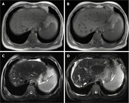
Figure 2 Magnetic resonance imaging images of biliary hamartomas on September 25, 2018.
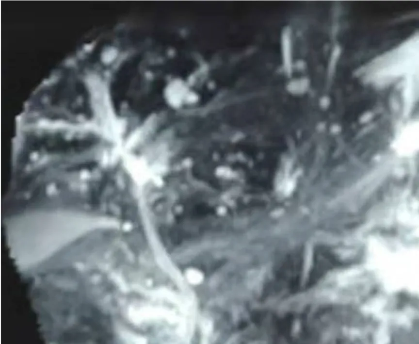
Figure 3 Magnetic resonance cholangiopancreatography image of biliary hamartomas on September 25, 2018.
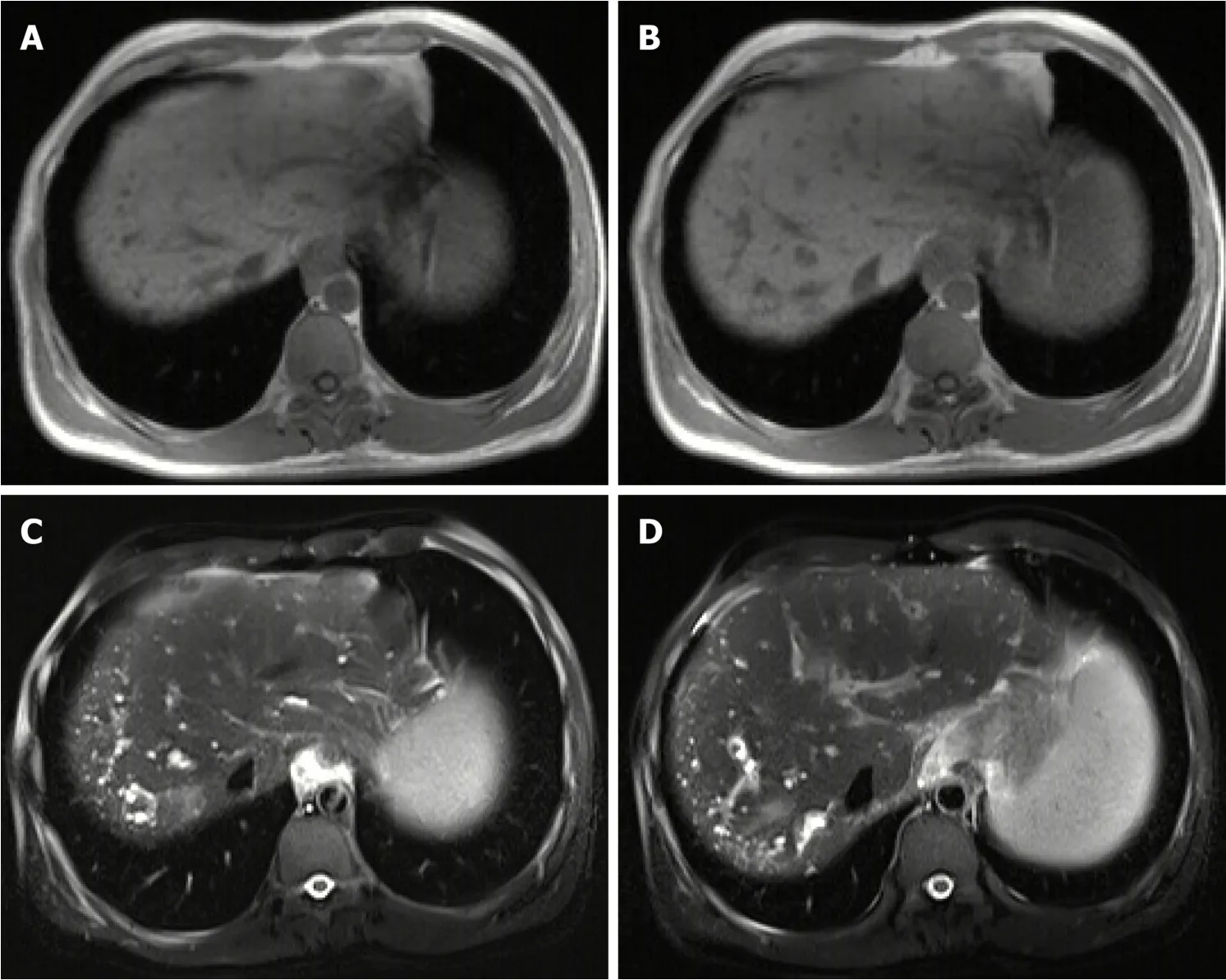
Figure 4 Magnetic resonance imaging images of biliary hamartomas on July 7, 2019.
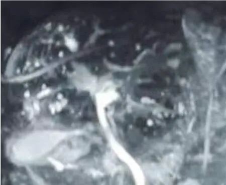
Figure 5 Magnetic resonance cholangiopancreatography image of biliary hamartomas on July 7, 2019.
 World Journal of Clinical Cases2020年9期
- World Journal of Clinical Cases的其它文章
- Rare primary lymphoepithelioma-like carcinoma of the renal pelvis
- Mesonephric adenocarcinoma of the uterine cervix with rare lung metastases:A case report and review of the literature
- Endoscopic ultrasonography elastography in the diagnosis of intrapancreatic ectopic spleen:A case report
- Giant ventral hernia simultaneously containing the spleen, a portion of the pancreas and the left hepatic lobe:A case report
- Application of curved ablation in liver cancer with special morphology or location:Report of two cases
- COVlD-19 managed with early non-invasive ventilation and a bundle pharmacotherapy:A case report