
Large carotid body tumor successfully resected in hybrid operating theatre:A case report
2019-03-14 04:11:20MengQiLiYanZhaoHuaiYuSunXinYuYang
World Journal of Clinical Cases 2019年16期
Meng-Qi Li, Yan Zhao, Huai-Yu Sun, Xin-Yu Yang
Abstract
Key words: Carotid body tumor; Paraganglioma; Hybrid operating theatre; Interventional embolization; Case report
INTRODUCTION
Carotid body tumor (CBT) is the most common paraganglioma in the head and neck[1].CBT typically originates from glomus caroticum in carotid bifurcation and consists of chemoreceptor cells of primitive neural crest origin[2-4].CBT can be categorized into three types:Sporadic, familial and hyperplastic[1].
Surgical resection is the standard treatment for CBT but carried substantial risk for large CBT, including cranial nerve injury and stroke[5].Intraoperative embolism has been used to successfully remove large CBT previously considered not resectable[6].Hybrid operating rooms are equipped with digital subtraction angiography (DSA)and near-infrared indocyanine green video angiography, and thus could readily identify severe complications in vascular surgery[7-9].
Here, we report a case of large CBT successfully resected after Onyx gel embolization of the feeding arteries in a hybrid operating theatre.
CASE PRESENTATION
Chief complaints
In July 2014, a 63-year-old right-handed woman presented with a progressively enlarging mass over the past five years and emerging numbness of the right hand and heel.
History of present illness
Patient’s symptoms started two months ago with recurrent numbness of the right hand and heel.
History of past illness
She had osteoarthritis of the knee for twenty years and hypertension for four years.
Physical examination
The mass was painless, non-moveable, and located between the right lower jaw and the right clavicle.No vascular murmur was heard over the mass upon auscultation.She had no dysphagia or voice hoarseness.No signs of hypoglossal neuropathy were detected.
Laboratory examinations
Laboratory blood tests were normal.
Imaging examinations
Magnetic resonance imaging (MRI) revealed a 130 mm × 60 mm × 70 mm mass with enhancement (Figure 1A and 1B).The mass spanned from the first to the seventh cervical vertebra, crossed over carotid bifurcation, and seemed to adhere to the right common carotid artery (CCA), right internal carotid artery (ICA), and right external carotid artery (ECA) (Figures 1C, D and Figure 2A).
FINAL DIAGNOSIS
The final diagnosis of the presented case is carotid body tumor; this is based on the clinical examination and postoperative pathology.
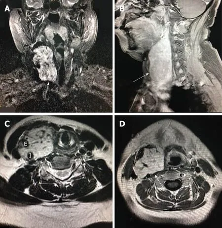
Figure 1 Enhanced magnetic resonance imaging.
TREATMENT
Surgical resection was conducted in a hybrid operating room, with a goal to completely resect the tumor (Figure 2B).DSA indicated that the right external carotid artery (ECA) was the major source of blood supply to the lesion (Figure 3A).A balloon test occlusion (BTO) was performed under general anesthesia.Upon angiography of the left ICA angiography, the right anterior cerebral artery and right middle cerebral artery were visualized.Twenty minutes after balloon occlusion, the patient’s neurological reflex was normal.BTO revealed poor compensation of the posterior communicating artery (PCoA) (Figure 3B and C).Onyx gel was used to embolize the feeding artery superior thyroid artery (STA) (Figure 3D).The ICA balloon was inflated to prevent gel entry into the right ICA.The procedure resulted in significant decrease in blood supply (Figure 3D) and the tumor began to shrink and harden.The balloon was deflated and removed from ICA, and surgical resection started.After isolation of the CBT, the blood supply from the right CCA, ICA, and ECA was blocked.The distal end of the right ECA was ligated, and the (including the part that adhered to the right ECA) was removed.At this point, the blood flow had been blocked for over 30 min.We restored blood flow of the carotid artery and put a vascular shunt between ICA and CCA.The part where the ICA was cut from the CCA was sutured.Intraoperative angiography at this time revealed patency of the ICA and its branches (Figure 4A).
OUTCOME AND FOLLOW-UP
Figure 4B shows the removed tumor.Post-operative pathological examination confirmed paraganglioma (Figure 4C).Two months after the surgery, numbness of the right hand and heel disappeared.Ultrasound examination showed that CCA and RICA were unobstructed (Figure 4D).Upon the last follow-up visit four years later,the patient had no recurrence and no any neurological abnormalities (Figure 4E).
DISCUSSION

Figure 2 Hypervascular carotid body tumor spans and remove of tumor.
To the best of our knowledge, the report represents the largest CBT that was successfully resected under interventional embolization in a hybrid operating room.
The CBT in the current case was very close to the base of the skull.As a result, the risk of significant blood loss and cranial nerve injuries during operation is high.A previous study showed that, for every 1-cm decrease in DTBOS, intraoperative blood loss (> 250 mL) increases by 1.8 times (95%CI:1.25-2.55), and the cranial nerve injury increases by 1.5 times (95%CI:1.19-1.92)[10].To reduce the intraoperative bleeding, we used Onyx gel to embolize the feeding artery.The right CCA, ICA and ECA were also blocked.We believe that these procedures are important for complete resection of the tumor.
Based on Shamblin's classification[4], CBT is classified into three types.Type I:Tumor is small and confined to the bifurcation of carotid artery, and there is little adhesion to the carotid artery; Type II:Tumor is large and partly surrounds the carotid artery with certain degree of adhesion to the carotid artery; and Type III:The tumor is large and completely envelops the carotid artery.CBT with higher Shamblin grade tends to have more severe neurological complications upon the surgery[11],mostly cranial nerve injuries[12,13].The tumor, in this case, was Shamblin type III, and unusually large, but neurological complications did not happen, possibly due to feeding artery embolization, blocking of the CCA, ICA and ECA, and experience of the surgeons.
It is noteworthy to point out that the management of large CBT should be individualized since the tumor could be located in varying positions and may invade different tissues[14,15].Also, an interdisciplinary approach is needed.
CONCLUSION
For large hypervascular CBT, embolization of the feeding artery in a hybrid operating theatre could be helpful.
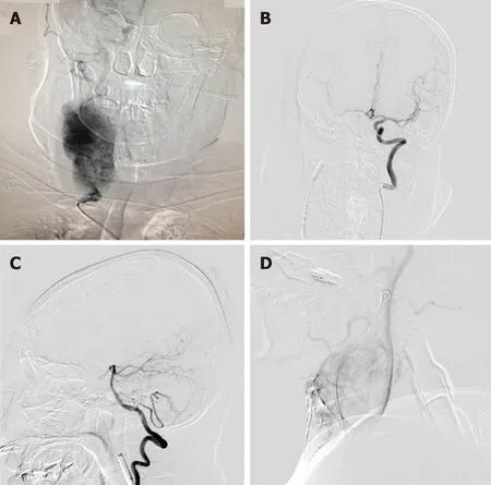
Figure 3 Preoperative angiography and balloon test occlusion.
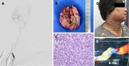
Figure 4 The tumor was completely removed and the right internal carotid artery flow was unobstructed.
ACKNOWLEDGEMENTS
We thank the index patient for the consent to publish this manuscript.
 World Journal of Clinical Cases2019年16期
- World Journal of Clinical Cases的其它文章
- Malignant syphilis accompanied with neurosyphilis in a malnourished patient:A case report
- Ex vivo revascularization of renal artery aneurysms in a patient with solitary kidney:A case report
- Pseudothrombus deposition accompanied with minimal change nephrotic syndrome and chronic kidney disease in a patient with Waldenstr?m's macroglobulinemia:A case report
- Hepatocellular carcinoma successfully treated with ALPPS and apatinib:A case report
- Treatment of invasive fungal disease:A case report
- Acute pancreatitis connected with hypercalcemia crisis in hyperparathyroidism:A case report