
Reduced microRNA-451 expression in eutopic endometrium contributes to the pathogenesis of endometriosis
2019-03-14 04:10:26ShanGaoShuangLiuZiMingGaoPengDengDanBoWang
World Journal of Clinical Cases 2019年16期
Shan Gao, Shuang Liu, Zi-Ming Gao, Peng Deng, Dan-Bo Wang
Abstract
Key words: Endometriosis; miR-451; Proliferation; Apoptosis; Pathogenesis
INTRODUCTION
Endometriosis (EMs) is a chronic and recurrent, but benign, disease in women of reproductive age, with a morbidity of approximately 10%.It is characterized by the presence of functional endometrial glands and stroma outside the uterine cavity[1,2].Typical symptoms of EMs include cyclic pelvic pain, dysmenorrhea, dyspareunia, and infertility.Previous studies have reported that EMs patients have a high risk of developing gynecological tumors and autoimmune disorders[3,4].Thus, EMs can cause severe psychological and physiological harm to those affected by it and imposes a substantial social burden[5,6].
Despite its high prevalence and incapacitating symptoms, the etiology of EMs is not clear.Evidence suggests that it is a multifactorial disease.Retrograde menstruation,immune system disorders, and genetic and environmental factors have been proposed as susceptibility factors for EMs[7-9].The susceptibility factor of retrograde menstruation proposed by Sampson is the most widely accepted[10].However, almost all women of reproductive age exhibit some degree of retrograde menstruation, and only 10% to 15% suffer from EMs[11,12].Recently, more evidence has emerged to support the theory that genetic changes in the eutopic endometrium may be the key molecular events in the pathogenesis of EMs[13].
MicroRNAs (miRNAs) are short noncoding RNA molecules that regulate genetic expression post-transcriptionally and are implicated in several biological processes,such as cell proliferation, differentiation, and apoptosis[14,15].Some miRNAs have been reported to be abnormally expressed in reproductive cancers[12,16,17], and miR-451 is of particular interest, as it acts as a tumor suppressor and is relevant to the poor prognosis of cancers.Aberrant miR-451 expression has been shown in eutopic and ectopic endometrial tissues; however, data regarding differences in miR-451 expression in the eutopic endometrium from healthy patients and those with EMs remain inconclusive[18,19].
In our study, we examined miR-451 expression in the eutopic endometrium of women with and without EMs and evaluated the role of miR-451 in cell proliferation.Finally, we predicted possible targets of miR-451 and the related signaling pathways.
MATERIALS AND METHODS
Tissue collection
Pathologic tissues were collected from patients with grade III cervical intraepithelial neoplasia, including 40 with EMs and 20 without.All 60 subjects underwent total hysterectomy at the Shengjing Hospital of China Medical University between 2009 and 2010.The EMs group included 2, 6, 20, and 12 cases at American Society for Reproductive Medicine (ASRM) stage I, II, III, and IV, respectively, of the disease.None of the patients had a history of endocrine, immune, or metabolic disorders and none had received any hormonal or antibiotic treatments within 3 mo prior to surgery.
Cell lines and culture
The ectopic and eutopic endometrial tissues were digested overnight at 37 °C with Dispase IV for 70 min and Dispase II for 50 min (Sigma, United States).After filtration through 100 and 400 mesh nylon screens, the obtained primary cells were rinsed in PBS and then cultured for 24 h in DMEM/F12 medium supplemented with 15% fetal bovine serum and antibiotics at 37 °C in an atmosphere containing 5% CO2.
Quantitative real-time PCR (qRT-PCR)
Total RNA was isolated from the EMs tissues using the TRIzol reagent.cDNA was synthesized from miR-451 using a TaqMan?miRNA Reverse Transcription Kit and used in a 1:5 dilution ratio for qRT-PCR, which was performed, using an miRNA Assays kit and Universal Master Mix following the kit protocols (Applied Biosystems).U6 was used as the endogenous control.Conditions of reverse transcription were as follows:16 °C for 30 min, 42 °C for 30 min, 85 °C for 5 min, and then holding at 4 °C.Conditions for qRT-PCR were as follows:enzyme activation at 95 °C for 10 min followed by 40 cycles of denaturation for 15 s at 95 °C and annealing and extension for 60 s at 60 °C.All experimental samples were run in triplicate and each qRT-PCR reaction was repeated at least two times.MiR-451 expression levels were calculated and analyzed using the 2?ΔΔCtrelative quantitation method.
Transfection
Using LipofectamineTM2000 reagent, miR-451 mimics and miR-451 inhibitors were transfected into EMs cells and normal endometrial cells, respectively.The oligonucleotide sequence of the miR-451 mimic is 5′-AAA CCG UUA CCA UUA CUG AGUU-3′, and its NC sequence is 5′-UUC UCC GAA CGU GUC ACG UTT-3′.The oligonucleotide sequence of the miR-451 inhibitor is 5′-AAC UCA GUA AUG GUA ACG GUUU-3′, and the sequence of the scrambled siRNA is 5′-CAG UAC UUU UGU GUA GUA CAA-3’.In addition, cells transfected with or without the empty vector were used as the control groups.All cells were incubated at 37 °C in an atmosphere containing 5% CO2for 24 to 96 h post transfection.
Cell proliferation analysis
Cellular proliferation analysis was performed using the Cell Counting Kit-8 (CCK-8)assay.After transfection with miR-451 mimics/inhibitors for 24h, 48h, 72h, and 96 h, 2× 10³cells were added to 96-well plates and incubated overnight at 37 °C in an atmosphere containing 5% CO2.Then, 10 μL of CCK-8 was added to each well(Beyotime Biotechnology).The cells were incubated for another 4 h at 37 °C in an atmosphere containing 5% CO2, and then cell viability was determined by measuring the optical density at 450 nm.
Flow cytometry to assess apoptosis
Annexin V-FITC/PI double-staining assays were performed for analysis of apoptosis.Cells were collected and suspended in PBS 24 h after transfection.Cells were then stained in 500 μL of binding buffer with 5 μL of each of annexin V-FITC and PI(KeyGen Biotech), incubated in the dark at room temperature for 5-15 min, and subjected to flow cytometric analysis to assess cellular apoptosis within 1 h.
Prediction of target genes and microarray data
Using miRDB (http://mirdb.org/miRDB/index.html) and miRcode(http://www.mircode.org/), we predicted the target genes of miR-451.Expression levels of the identified targeted genes were determined by analyzing the GSE7846 gene profile from the GEO database (https://www.ncbi.nlm.nih.gov/geo/).This dataset includes the expression data of endometrial cells derived from patients with EMs (ectopic group) and without EMs (normal group).We screened the differentially expressed genes with aP-value < 0.01 and an adjustedP-value < 0.01 between the EMs and control groups.
Statistical analysis
Data are expressed as the mean ± SEM.Statistical comparisons between groups were determined using thet-test andχ2test.Statistical significance was defined asP< 0.05.Analyses were performed using the R 3.4.2 and SPSS 22.0 software.
Ethics approval and informed consent
This study was approved by the China Medical University Research Ethics Committee according to the Helsinki Declaration, and written informed consent was obtained from each study participant.
RESULTS
MiR-451 expression is reduced in eutopic tissues and cell lines derived from EMs patients compared to the controls
qRT-PCR was performed to quantitatively analyze the expression levels of miR-451 in eutopic tissues from the EMs and control groups.As shown in Figure 1A, we observed a significant reduction in miR-451 expression in the EMs group compared to the control group (EMs, 0.22 ± 0.06; control, 1.12 ± 0.11,P< 0.01).Consistent with the tissue results, miR-451 expression in cells was significantly lower in the EMs group than in the control group (Figure 1B).The correlation of miR-451 expression levels with ASRM stage was then analyzed, as shown in Table 1.No significant association was found between miR-451 expression and ASRM stage (P> 0.05).
MiR-451 mimic inhibits cell proliferation and induces apoptosis in EMs eutopic cells
MiR-451 levels in eutopic cells transfected with miR-451 mimic were higher than those in the non-transfected control and scrambled mimic oligomer groups (miR-451 mimic,1.33 ± 0.28; control, 0.25 ± 0.06; scrambled, 0.32 ± 0.09,P< 0.01) (Figure 2A).CCK-8 assay results showed that transfection with miR-451 mimic suppressed the proliferation rate of EMs cells (Figure 2B).To investigate whether the reduced cell proliferation resulted from apoptosis, we evaluated the effect of miR-451 mimic on cellular apoptosis using flow cytometry.MiR-451 mimic induced early apoptosis in a larger number of cells compared to scrambled oligonucleotides, and this difference was statistically significant (P< 0.01) (Figure 2C and 2D).Thus, overexpression of miR-451 in EMs cells induces apoptosis and inhibits cell proliferation.
Transfection of miR-451 siRNA into control eutopic cells promotes cell proliferation and inhibits cell apoptosis
As shown in Figure 3A, miR-451 expression was significantly attenuated in the siRNA-transfected group compared to the non-transfected and scrambled mimic oligomer groups (miR-451 siRNA, 0.41 ± 0.14; control, 1.23 ± 0.08; scrambled, 1.06 ±0.06,P< 0.01).Additionally, the proliferation ability of miR-451 siRNA-transfected cells was greater than that of the other two groups (Figure 3B).We used flow cytometric assay to evaluate the effect of miR-451 siRNA transfection on apoptosis.Our results showed that the proportion of early apoptotic cells was significantly lower in the miR-451 siRNA group compared to that in the scrambled group (P< 0.01)(Figure 3C and 3D).These results indicate that, in eutopic cells, miR-451 reduces apoptosis and increases cell proliferation.
Prediction of miR-451 target genes
Using the miRDB and miRcode miRNA target prediction databases, we identified a total of 12 genes targeted by miR-451, namely,OSR1, MEX3C, CUX2, ZNF644,TBC1D9B, DCAF5, CDKN2B, TTN, YWHAZ, CDKN2D, EIF2AK3, andTBX1(Figure 4A).As shown in Figure 4B, among the targeted genes, the expression levels ofOSR1,YWHAZ,TTN, andCDKN2Dwere significantly different between the two groups according toGSE7846(P< 0.05, adj.P< 0.05).The logFC values ofOSR1,YWHAZ,TTN, andCDKN2Dwere 0.76, 0.43, 0.33, and 0.63, respectively.Furthermore,according to the pathway analysis data in the Kyoto Encyclopedia of Genes and Genomes,YWHAZandCDKN2Dmay have important roles in the cell cycle in EMs(Figure 4C).
DISCUSSION

Table 1 Comparison of miR-451 expression in patients at different american society for reproductive medicine stages
In this study, qRT-PCR analysis of eutopic endometrial tissues and cells showed that miR-451 was significantly downregulated in patients with EMs compared to normal controls (P= 0.011).Although we did not observe a significant association between miR-451 expression and the ASRM stage of EMs, ectopic overexpression of miR-451 in eutopic cells in EMs was shown to be associated with reduced cell proliferation and increased apoptosis.Conversely, siRNA-mediated knockdown of miR-451 promoted the proliferation and reduced the apoptosis of eutopic cells.
The “eutopic endometrium determinism” theory suggests that the occurrence of EMs is mainly dependent on the characteristics of eutopic endometrial lesions, and retrograde menstruation may act as a precipitating factor.Thus, genetic dysregulation in the endometrium is crucial in the pathogenesis of EMs.Identifying differentially expressed genes between patients with and without EMs would serve as a minimally invasive method to diagnose EMs and evaluate the risk of recurrence.For example,Mahdianet al[20]reported thatMIF,CD74, andCOX-2are essential in inflammation and endometrium reconstruction during the menstrual cycle, and increased expression of these genes is a molecular biomarker for the development and pathophysiology of EMs.In addition, Sapkotaet al[21]also identified five novel loci(CCDC170,FN1,SYNE1,ESR1, andFSHB) and nineteen independent single nucleotide polymorphisms that are significantly associated with the risk of EMs.
Furthermore, miRNAs regulate the expression of target genes and key cellular processes in EMs.In 2009, Burneyet al[14]reported the downregulation of the miR-9 and miR-34 miRNA families in eutopic cells in the setting of EMs, and this downregulation is closely related to progesterone resistance in early secretory endometrium.Laudanskiet al[22]showed that miR-483-5p and miR-629-3p are downregulated in EMs, and this is associated with inflammation.Moreover, miR-21 was shown to be significantly upregulated in severe EMs (stage III/IV) compared to mild EMs (stage I/II)[23].Notably, miR-451 has been established as a tumor-suppressor gene in gastric, colorectal, bladder, and non-small cell lung carcinomas[24], and it was also shown to be downregulated in ovarian cancer compared to its concurrent EMs[25].In addition, Nothnicket al[26]showed that deficiency of miR-451 regulates fibrinogen alpha chain and reduces endometrial implantation in a mouse model.Similarly, we found that miR-451 was downregulated in the eutopic endometrium in EMs compared to normal controls in studies involving both tissues and cells.
Using miRNA target-predicting databases, we identified 12 potential target genes of miR-451 and analyzed their expression levels according to theGSE7846dataset.Finally, a total of four genes,YWHAZ,OSR1,TTN, andCDKN2D, were selected for further analysis.Among these target genes of miR-451,YWHAZhas previously been shown to be overexpressed in tissues in EMs[19,27].Joshiet al[19]reported that miR-451 regulatesYWHAZexpression and promotes proliferation of eutopic cells in baboons with EMs[19].However, the roles ofOSR1,TTN, andCDKN2Din EMs have not been reported until now.Published reports suggest thatOSR1inhibits proliferation and induces cellular apoptosis by acting on the WNK and NF-κB pathways, andOSR1is dramatically downregulated in several carcinomas[28-30].In addition,CDKN2Dhas been shown to be involved in carcinogenesis and has been identified in gynecological cancers.This gene may be regulated by miR-451 in esophageal carcinoma cell lines[31].Thus, our study provides several novel therapeutic targets for EMs.
Notably, most studies on EMs have only focused on identifying differences between ectopic lesions and eutopic endometrium.For example, Grahamet al[18]reported that miR-451 is overexpressed in ectopic lesions compared to eutopic lesions and reduces cell survival by regulatingMIF.In this study, we found significant differences in miR-451 expression in the eutopic endometrium of patients with and without EMs, which effectively supports the “eutopic endometrium determinism”theory.Furthermore, we identified four potential target genes of miR-451 by bioinformatics analysis and analyzed their downstream pathways.
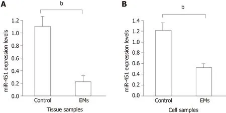
Figure 1 Expression of miR-451 in eutopic tissues and cell lines.
Our study has two limitations.First, the number of included patients was relatively small.Second, thein silico-predicted targets of miR-451 need to be validated through experiments, such as 3′-UTR luciferase reporter assays.However, we believe that our results indicate a novel role of miR-451 in EMs and support several potential biomarkers in the form of miR-451 targets that may be used for future clinical diagnosis and therapy of this disease.
In conclusion, miR-451 is a novel biomarker for EMs and is downregulated in the eutopic endometrium.YWHAZ,OSR1,TTN, andCDKN2Dare potential target genes of miR-451 and may have important roles in the pathogenesis of EMs.

Figure 2 Transfection with miR-451 mimic inhibits cell proliferation by inducing the apoptosis of eutopic cells in endometriosis.

Figure 3 Transfection with miR-451 siRNA decreases apoptosis and promotes cell proliferation in control eutopic cells.

Figure 4 Results of bioinformatics analysis of miR-451 target genes according to the GSE7846 dataset.
ARTICLE HIGHLIGHTS
Research background
Despite the high prevalence of endometriosis (EMs), its etiology is unclear.
Research motivation
MiR-451 acts as a tumor suppressor and is relevant to the poor prognosis of cancers.
Research objectives
To evaluate the expression levels and role of miR-451 in the eutopic endometrium and predict possible targets of miR-451 and related signaling pathways.
Research methods
Quantitative real-time PCR was used to evaluate miR-451 expression.Cell Counting Kit-8 and flow cytometric assays were performed to determine cell proliferation and survival rates.
Research results
MiR-451 was downregulated in the eutopic endometrium and related with EMs cell proliferation and apoptosis.YWHAZ,OSR1,TTN, andCDKN2Dwere identified as potential target genes of miR-451.
Research conclusions
Reduced miR-451 expression in the eutopic endometrium contributes to the pathogenesis of EMs by promoting cell proliferation and reducing apoptosis.
Research perspectives
MiR-451 is a novel biomarker for EMs.YWHAZ,OSR1,TTN, andCDKN2Dare potential target genes of miR-451 and may have key roles in this disease.
 World Journal of Clinical Cases2019年16期
- World Journal of Clinical Cases的其它文章
- Malignant syphilis accompanied with neurosyphilis in a malnourished patient:A case report
- Ex vivo revascularization of renal artery aneurysms in a patient with solitary kidney:A case report
- Pseudothrombus deposition accompanied with minimal change nephrotic syndrome and chronic kidney disease in a patient with Waldenstr?m's macroglobulinemia:A case report
- Hepatocellular carcinoma successfully treated with ALPPS and apatinib:A case report
- Treatment of invasive fungal disease:A case report
- Acute pancreatitis connected with hypercalcemia crisis in hyperparathyroidism:A case report