
Chronic atrophic endometritis and pyometra in a ferret: A case report
2019-02-14 05:52:42AntonLazarinovAntonovRadostinStefanovSimeonovKoychoPetkovKoev
Anton Lazarinov Antonov, Radostin Stefanov Simeonov, Koycho Petkov Koev
1Department of Obstetrics, Reproduction and Reproductive Disorders, Faculty of Veterinary Medicine, Trakia University, Stara Zagora, 6000, Bulgaria
2Department of General and Clinical Pathology, Faculty of Veterinary Medicine, Trakia University, Stara Zagora, 6000, Bulgaria
3Department of Veterinary Microbiology, Infectious and Parasitic Diseases, Faculty of Veterinary Medicine, Trakia University, Stara Zagora, 6000, Bulgaria
Keywords:Ferret Uterus Endometritis Pyometra
ABSTRACT The aim of this report was to describe a clinical case of chronic atrophic endometritis as a complication of cystic endometrial hyperplasia-pyometra complex in a non-spayed ferret.The ferret was presented with a slight abdominal distension and odorless purulent vulvar discharge after unsuccessful medical treatment with enrofloxacine and aglepristone 2 months ago in another clinic. Ultrasonography revealed enlarged uterine horns filled with fluid and blood laboratory analysis showed anaemia and leukocytosis, so diagnosis of pyometra was made. Laparotomy and ovariohysterectomy were performed. Histopathological and microbiological examination of the uterus revealed the presence of purulent atrophic endometritis caused by Staphylococcus spp. In conclusion, this is a very rare case of endometrial atrophia after chronic uterine inflammation in a ferret.
1. Introduction
Pyometra in ferrets (Mustela putorius furo) has been previously reported as a complex with cystic endometrial hyperplasia (CEH)[1,2]. The uterine inflammation is a result of invasion most often by Escherichia coli entering through the vagina[3]. CEH is suspected to be one of the predisposing factors for the development of infection because of the cystic distention of uterine glands, fibrosis,accumulation of intraluminal fluids, development of crypts and cysts where bacteria can proliferate[4].
CEH-pyometra complex is associated with increased levels or imbalance of ovarian steroids[3]. Progesterone stimulates endometrial gland secretion, supresses the immune responses,diminishes myometrial activity, causes functional closure of the cervix which inhibits the drainage of uterine fluids and provides a suitable environment for bacterial growth[5]. Estrogen increases the number of progesterone receptors and persistently high levels may lead to inappropriate proliferation of the endometrium[6]. The repeated and prolonged response to steroids may lead to endometrial changes like hydrometra, mucometra and if bacterial invasion occurs to pyometra[7,8].
In ferrets, CEH-pyometra complex is associated with the presence of adrenal gland disease, remnant ovarian tissue and ovarian cancer[2,3].
In our opinion, it is a very rare report of a case of chronic atrophic endometritis as a complication of pyometra in a ferret.
2. Case description
A 5-year-old multiparous ferret, weighting 0.81 kg, was presented to the Small Animal Clinic of the Faculty of Veterinary Medicine,Trakia University in Stara Zagora, Bulgaria, with signs of slight abdominal distension and odorless purulent vulvar discharge. The owner reported that these signs spontaneously started 2 months ago and the ferret was brought to another clinic where the veterinarian prescribed medical treatment with enrofloxacine and aglepristone for 5 consecutive days.
On physical examination, there were no changes in the general condition of the animal. Rectal body temperature was 38.8 ℃, heart rate was 200/min, and respiratory rate was 39/min. The colour of visible mucosa coats was rose-red.
At the inspection of the external genitalia it was found that the vulva was oedematous and with odorless purulent discharge (Figure 1). Radiographically a soft tissue mass was found into the abdomen(Figure 2). Transabdominal ultrasonography (Mindray DC-6 Vet,China, 6.5 MHz convex transducer) revealed fluid-filled uterine horns (Figure 3). The urinary bladder was found in its normal anatomic location.
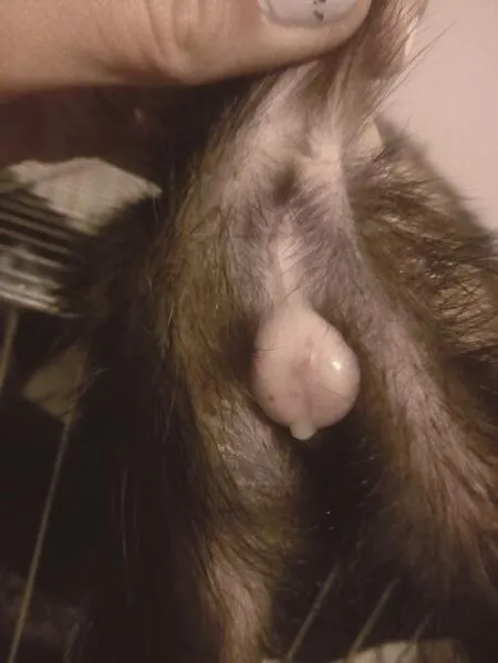
Figure 1. Vulva with purulent discharge of ferret.
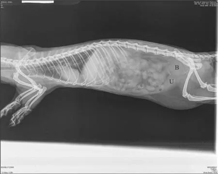
Figure 2. Radiography: lateral view.
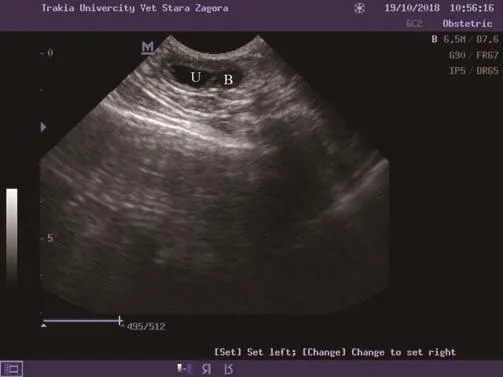
Figure 3. Ultrasonographic image of fluid-filled uterus of ferret.
To determine complete blood cell counts and biochemical parameters, blood samples (about 1 mL) were collected by venipuncture of the cephalic vein. Complete blood cell counts were assayed using an automated haematological analyser BC-2800 Vet(Mindray, China), and blood biochemical parameters were tested using an automated biochemical analyser BS 120 (Mindray, China).Blood laboratory analysis showed the presence of anaemia and leukocytosis (Table 1).
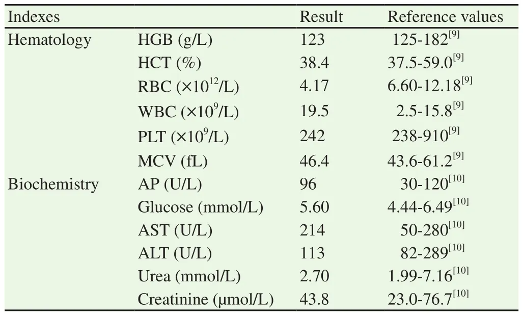
Table 1 Hematological and blood biochemistry analysis results.
The ferret was diagnosed with pyometra, so a decision for laparotomy was taken. After aseptic preparation of the abdominal region, the ferret was premedicated intramuscularly with 0.5 mg/kg butorphanol (Butomidor; Richter pharma; Austria). Anaesthesia was induced by mask and after endotracheal intubation maintained with isoflurane (TerrellTM; Minrad Inc.; USA). The ferret was placed in a dorsal recumbency and a caudal median laparotomy was performed.Gross examination of the reproductive tract revealed enlarged uterine horns (Figure 4). An ovariohysterectomy was performed.The abdominal cavity was closed with cross stitch pattern using USP 3/0 polyglycolic acid absorbable sutures (Marlin; Catgut GmbH;Markneukirchen), and the skin was sutured with simple interrupted non-absorbable sutures USP 2/0 (Vitalon; Dr Hammer & Co.GmbH; Hamburg). After the operation, incisions of the uterine horns were made and macroscopically there was a significant thickening of the uterine wall and the lumen of the uterus was filled with small amount of greenish substance (Figure 5). Postoperative treatment included antibiotic – 15 mg/kg amoxicillin-clavulanic acid (Synulox RTU; Zoetis; Belgium), subcutaneously for 5 d.
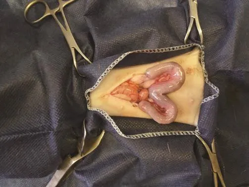
Figure 4. Intraoperative appearance of uterus of ferret.
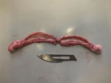
Figure 5. Internal genitalia of ferret after ovariohysterectomy and incision of uterine horns.
Microbiological evaluation of the uterine fluid detected the presence ofStaphylococcusspp. sensitive to amoxicillin-clavulanic acid, cefquinone, cefalexin and sulphonamides and resistant to enrofloxacin, penicillin, ampicillin and lincomycin.
The excised uterus was submitted for histopathology. The specimens were fixed in 10% neutral formalin and processed routinely. The 4 μm cross sections were stained with haematoxylineosin. Multiple uterine thickenings were detected (Figure 6). The cluster of fibroblasts and fibrocytes was observed in the lamina propria, leading to fibrosis and sclerosis of the endometrium (Figure 7). Due to compression from the connective tissue, the uterine glands were atrophied (chronic atrophic endometritis). The epithelial cells of the remaining single glands were pyknotic, and the cytoplasm was grainy and enlightened. Surface epithelial cells were desquamated.Skin sutures were removed after 10 d. The post-operative examination showed that the ferret was in a good condition.
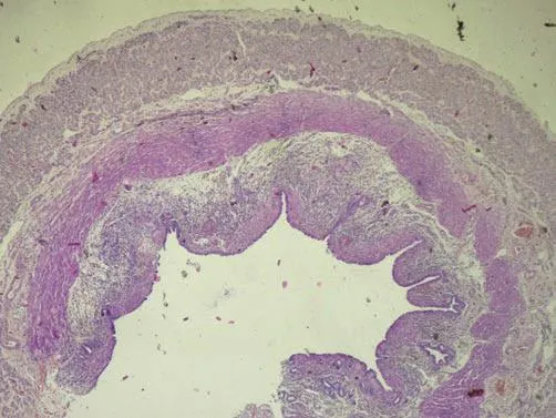
Figure 6. Uterus with multiple thickening of wall.
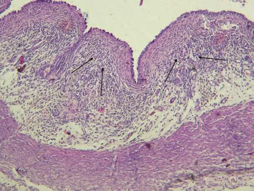
Figure 7. Uterus.
3. Discussion
The prevalence of CEH-pyometra complex in ferrets has not yet been determined[2]. Desexing in this species is performed routinely between the age of 6 weeks in the USA and several months in Europe[7,11]. The previously reported cases described CEH-pyometra complex after ovariectomy or ovariohysterectomy associated with ovarian remnant, ovarian cancer and adrenal disease[2]. Our case was an intact female which had one litter of 4 three years before the occurrence of uterine inflammation. Probably the prolonged estrus season may be the reason for the disease of uterus. The inflammation was long lasting (2 months) and after unsuccessful conservative therapy, so a decision for surgical treatment was done. The reason for this could be found in the microbial resistance of the isolated culture ofStaphylococcusspp. from the uterine content to enrofloxacine.
The most interesting and unique fact was that histopathologically the atrophied uterine glands were found, so the ferret was diagnosed with chronic atrophic endometritis. In previously described cases, the animals had cystic endometrial hyperplasia with increased number of endometrial glands because of the estradiol stimulation[1,2,7,11].The reason for the local degenerative changes within uterine tissues in our case could be found in the prolonged inflammation process.
This is a very rare report of such a degenerative inflammation uterine process in ferrets. In cases of prolongation and absence of success of the medical treatment, we recommend a complete ovariohysterectomy. Early age desexing of the female ferrets may be used in order to prevent the diseases of the reproductive organs.
Conflict of interest statement
All the authors declare that there is no conflict of interest.
 Asian Pacific Journal of Reproduction2019年1期
- Asian Pacific Journal of Reproduction的其它文章
- Infertility in China: Culture, society and a need for fertility counselling
- Total segmental aplasia of uterus body in bitch
- Comparison of transvaginal cervical length and modified Bishop’s score as predictors for labor induction in nulliparous women
- Value of α-fetoprotein,β-HCG, inhibin A, and UE3 at second trimester for early screening of preeclampsia
- Blood indicators of dry cows before and after administration of a drug STEMB
- Effect of butylated hydroxytoluene on quality of pre-frozen and frozen buffalo semen