
Recovery of injured fornical crura following neurosurgical operation of a brain tumor: a case report
2016-12-02 07:05:45SungHoJang,YouSungSeo
中國神經(jīng)再生研究(英文版) 2016年5期
IMAGING IN NEURAL REGENERATION
Recovery of injured fornical crura following neurosurgical operation of a brain tumor: a case report
The fornix, which is involved in transfer of episodic memory, is an important structure of the Papez circuit between the medial diencephalon and the medial temporal lobe. Many studies using DTI have reported on injury of the fornix in patients with brain injury (Nakayama et al., 2006; Sugiyama et al., 2007; Wang et al., 2008; Chang et al., 2010). However, only a few studies on recovery of an injured fornix in patients with brain injury, including traumatic brain injury and stroke, have been reported (Yeo et al., 2011; Yeo and Jang, 2013a, b). In this study, using follow up DTT images, we reported on a patient who showed recovery of injured fornical crura following a neurosurgical operation for a brain tumor.
A 48-year-old female patient underwent craniotomy and a navigator assisted removal of craniopharyngioma at the university hospital. Brain MRIs showed a cystic mass at the suprasellar space before the operation, and a lesion and hematoma in the body of the corpus callosum and the lateral ventricle 1 day after the operation (Figure 1A). According to pathology, the patient was diagnosed suffering from a craniopharyangioma. The patient had shown severe memory impairment since the operation and underwent neuropsychological evaluations at 6 weeks after onset: Memory Assessment Scale [MAS, global memory: 58 (<1%ile), short term memory: 61(< 1%ile), verbal memory: 58(<1%ile), and visual memory: 60(< 1%ile)], Mini-Mental State Examination (MMSE: 13). She had undergone comprehensive rehabilitation, including cognitive therapy, until 20 months after onset. She showed mild improvement in memory impairment at 20 months after onset: MAS [global memory: 61 (> 1%ile), short term memory: 101 (53%ile), verbal memory: 67 (1%ile), and visual memory: 70 (2%ile)], MMSE: 24.
DTI was performed three times (2, 6 and 20 months after onset) using a 6-channel head coil on a 1.5T Philips Gyroscan Intera. Fiber tracking was based on the fiber assignment continuous tracking (FACT) algorithm implemented within the DTI task card software. For reconstruction of the fornix, the first region of interest (ROI) was placed at the junction between the body and column of the fornix on a coronal image of the color map. The second ROIs were placed on each side of the crus of the fornix on a coronal image of the color map, respectively (Yeo and Jang, 2013b). On 2-month DTT images, discontinuations were observed in both fornical crura (Figure 1B). A collateral branch from the end of the right fornical body was observed, which ended in the right temporal lobe via the splenium of the corpus callosum. On 6-month DTT images, fornical crura emerged from the end of the fornical body on both sides; in contrast, the right collateral branch was shortened and a collateral branch emerged from the left fornical body. On 20-month DTT images, the fornical crura were elongated to the medial temporal lobe on both sides and the collateral branches had disappeared on both sides.
In the current study, we investigated changes in DTT findings of injured fornical crura following a neurosurgical operation in a patient with a brain tumor. After removal of the tumor, a lesion and hematoma were observed in the body of the corpus callosum and the lateral ventricle. We observed discontinuations of the bilateral fornix crura on 2-month DTT images. Based on previous studies demonstrating the vulnerability of the fornix to intraventricular hemorrhage and lesion in the corpus callosum, the fornix injury in this patient was ascribed to the intraventricular hemorrhage and neurosurgical operation through the body of the corpus callosum (Chang et al., 2010; Yeo et al., 2011). We observed two kinds of changes of injured fornical crura on three follow up DTT images: 1) recovery of discontinued fornical crura on both sides, consequently, theends of injured fornical crura were connected to the medial temporal lobe on both sides on 20-month DTT images, and 2) the collateral branches via the splenium of the corpus callosum from the injured fornical body disappeared with the recovery of injured fornical crura. Previous studies have reported that collateral branches via the splenium of the corpus callosum result from development of compensatory neural tracts following injury of the fornical crus (Yeo and Jang, 2013a, b; Lee et al., 2014). Therefore, we believe that the compensatory collateral branches via the corpus callosum had disappeared along with the recovery of the injured fornical crura. The improvement of impaired memory in the visual and verbal observed in this patient for 20 months may be additional evidence of the recovery of injured fornical crura in this patient.
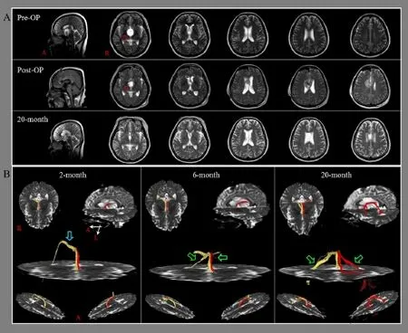
Figure 1 Brain magnetic resonance and diffusion tensor tractography (DTT) images of the fornical crura in a patient with a brain tumor after neurosurgical operation.
Since introduction of DTI, a few studies have reported on the recovery of an injured fornix in patients with brain injury (Yeo and Jang, 2013a, b; Lee et al., 2014). In 2013, Yeo and Jang (2013b) reported on a patient who showed compensatory neural tracts (an abnormal neural tract originating from an injured fornical crus passed through the splenium of the corpus callosum to connect to the medial temporal lobe and another abnormal neural tract originating from the fornical column was connected to the medial temporal lobe) after head trauma resulting in bilateral injury of the fornical crura. Yeo and Jang (2013a) also reported on a patient who underwent coiling for a ruptured anterior communicating cerebral artery aneurysm and conservative management for subarachnoid and intraventricular haemorrahage. They showed that the end of the discontinued fornical body was connected to the splenium of the corpus callosum and then branched to the medial temporal lobe and thalamus. Lee and Jang (2014) recently reported on the change of an injured fornix in a patient with traumatic axonal injury. One branch from the injured fornical body was connected to the medial temporal lobe via the splenium of the corpus callosum, and two branches from both fornical columns were connected to the medial temporal lobes respectively. As a result, to the best of our knowledge, this is the first study to demonstrate the recovery of injured fornical crura in a patient with brain injury. However, limitation of DTI should be considered (Yamada et al., 2009). DTI may underestimate or overestimate the fiber tracts. In addition, fiber complexity and crossing can prevent full reflection of the underlying fiber architecture by DTI (Yamada et al., 2009).
In conclusion, we reported on a patient who showed recovery of injured fornical crura following a neurosurgical operation for a brain tumor. This finding appears to suggest a mechanism for recovery of injured fornical crura. Because it is based on a single case report, this study is limited. Larger-scale complementary studies needed to be conducted.
This work was supported by the National Research Foundation (NRF) of Korea Grant funded by the Korean Government (MSIP), No. 2015R1A2A2A01004073.
Sung Ho Jang, You Sung Seo*
Department of Physical Medicine and Rehabilitation, College of Medicine, Yeungnam University, Daemyungdong, Namku, Daegu, Republic of Korea
*Correspondence to: You Sung Seo, M.S., yousung1008@daum.net.
Accepted: 2016-02-04
orcid: 0000-0002-7480-3071 (You Sung Seo)
Chang MC, Kim SH, Kim OL, Bai DS, Jang SH (2010) The relation between fornix injury and memory impairment in patients with diffuse axonal injury: a diffusion tensor imaging study. NeuroRehabilitation 26:347-353.
Concha L, Gross DW, Beaulieu C (2005) Diffusion tensor tractography of the limbic system. Am J Neuroradiol 26:2267-2274.
Lee HD, Jang SH (2014) Changes of an injured fornix in a patient with mild traumatic brain injury: Diffusion tensor tractography follow-up study. Brain Inj 28:1485-1488.
Nakayama N, Okumura A, Shinoda J, Yasokawa YT, Miwa K, Yoshimura SI, Iwama T (2006) Evidence for white matter disruption in traumatic brain injury without macroscopic lesions. J Neurol Neurosurg Psychiatry 77: 850-855.
Sugiyama K, Kondo T, Higano S, Endo M, Watanabe H, Shindo K, Izumi S (2007) Diffusion tensor imaging fiber tractography for evaluating diffuse axonal injury. Brain Inj 21:413-419.
Wang JY, Bakhadirov K, Devous MD Sr, Abdi H, McColl R, Moore C, Marquez de la Plata CD, Ding K, Whittemore A, Babcock E, Rickbeil T, Dobervich J, Kroll D, Dao B, Mohindra N, Madden CJ, Diaz-Arrastia R (2008) Diffusion tensor tractography of traumatic diffuse axonal injury. Arch Neurol 65:619-626.
Yamada K (2009) Diffusion tensor tractography should be used with caution. Proc Natl Acad Sci U S A 106:E14.
Yeo SS, Choi BY, Chang CH, Jung YJ, Ahn SH, Son SM, Byun WM, Jang SH (2011) Periventricular white matter injury by primary intraventricular hemorrhage: a diffusion tensor imaging study. Eur Neurol 66:235-241.
Yeo SS, Jang SH (2013a) Recovery of an injured fornix in a stroke patient. J Rehabil Med 45:1078-1080.
Yeo SS, Jang SH (2013b) Neural reorganization following bilateral injury of the fornix crus in a patient with traumatic brain injury. J Rehabil Med 45:595-598.
Copyedited by Lin S, Petrosyan T, Shan J, Li CH, Song LP, Zhao M
10.4103/1673-5374.182714 http∶//www.nrronline.org/
How to cite this article: Jang SH, Seo YS (2016) Recovery of injured fornical crura following neurosurgical operation of a brain tumor: a case report. Neural Regen Res 11(5):854-855.
- 中國神經(jīng)再生研究(英文版)的其它文章
- Gender difference in the neuroprotective effect of rat bone marrow mesenchymal cells against hypoxiainduced apoptosis of retinal ganglion cells
- Vitamin B complex and vitamin B12levels after peripheral nerve injury
- Methylprednisolone microsphere sustained-release membrane inhibits scar formation at the site of peripheral nerve lesion
- A self-made, low-cost infrared system for evaluating the sciatic functional index in mice
- Methylprednisolone exerts neuroprotective effects by regulating autophagy and apoptosis
- Repetitive magnetic stimulation affects the microenvironment of nerve regeneration and evoked potentials after spinal cord injury