
Extraskeletal Chondroma in the Popliteal Region: A Case Report
2015-11-18 01:19:26BingyanMao
Chinese Medical Sciences Journal 2015年4期
Bing-yan Mao*
Department of Orthopaedic Surgery, Shimen Hospital of Changsha Medical University, Changde 415300, Hunan, China
Extraskeletal Chondroma in the Popliteal Region: A Case Report
Bing-yan Mao*
Department of Orthopaedic Surgery, Shimen Hospital of Changsha Medical University, Changde 415300, Hunan, China
extraskeletal chondroma; popliteal region
Chin Med Sci J 2015; 30(4):270-272
E XTRASKELETAL chondroma is a rare benign tumor that usually develops in the soft tissue, which commonly occurs in the limbs of adults and generally presents as painless masses without the surrounding tissues involvement. The diagnosis mainly depends on computed tomography, magnetic resonance imaging (MRI) examination, and postoperative pathological examination. This paper reports a case of extraskeletal chondroma in the popliteal region.
CASE DESCRIPTION
A 55-year-old woman was referred to our hospital who complained of a mass in the left popliteal region. She had suffered from pain for three years and limited mobility in her left leg for six months.
The physical examination showed a palpable, hard, mobile mass with clear boundary in the left popliteal fossa and no palpable lymph nodes. The popliteal area was slightly swollen with the normal local skin temperature. The localized tenderness and skin irritation was observed. She denied radiating pain in the left leg. There was no vascularmurmur. She presented with an aggravated pain with flexion and slight limitation of movement in the left knee.
Laboratory examination revealed the routine blood test, serum alkaline phosphatase level, erythrocyte sedimentation rate, rheumatoid factor, and high-sensitivity C-reactive protein were normal. X ray photograph showed the bone structure in the knee was normal. MRI displayed a cystic lesion in the posterior part of the left knee joint accompanied with effusion, degenerative changes of both the anterior and posterior horns of the medial meniscus, and bone marrow damage (Fig. 1). Therefore, the patient was primarily diagnosed with a mass of undetermined origin in the left popliteal region.
The lesion was fully resected using standard surgical approach under epidural anesthesia. The mass was completely encapsulated with no obvious connection to the peripheral tissues and osseous structure of knee joint or joint capsule, measuring approximately 4.0 cm × 3.7 cm × 3.0 cm (Fig. 2A). The boundary of the specimen was clear; mucoid degeneration and cystic degeneration appeared in the center of lesion (Fig. 2B). Histopathological examination revealed the tumor was composed of mature hyaline cartilage and fibrocartilage with different levels of mucoid and cystic changes. Chondroma cells exhibited polymorphic without nuclear atypia and mitosis (Fig. 2C, 2D). Pathologicaldiagnosis was extraskeletal chondroma in the left popliteal region.
After surgery the local deformity disappeared, the pain was greatly released, and flexion and extension function of the knee restored normal. No local recurrence was found in the six-month of postoperative follow-up.
DISCUSSION
Extraskeletal chondroma is a rare benign tumor that predominantly occurs in the soft tissue of the limbs,1,2accounting for only 1.5% benign soft-tissue tumors. The tumor is usually present as a single, painless mass. Generally, the lesion grows slowly and its size seldom exceeds 3.0 cm in diameter. It is often seen at the ages of 30-60 years with an equal sex distribution,3,4when the tumor has increased big enough to cause local deformity or intermittent pain. The underlying etiology is unclear. It has been manifested that extraskeletal chondroma might arise from residual embryonal tissue or from undifferentiated mesenchymal stem cells.4,5
The histopathological examination showed that the extraskeletal chondroma was completely composed of mature hyaline cartilage and fibrocartilage, which had no connection with the bone or periosteum tissues and had different levels of calcification, mucoid and cystic changes. Cellular polymorphism with rare mitotic figures and without atypism is the typical pattern of tumor cells. The peripheral zones of a few lobular cartilages have a high density of spindle-shaped stromal cells, which had no perinuclear halos or lacuna-shaped structures. Immunohistochemistrically, the tumor cells were positive for S-100 protein and vimentin, but were negative for the epithelial and myoepithelial markers.6,7
To our knowledge, the mucoid and cystic changes in extraskeletal chondroma have rarely been reported. For our case, the mucoid and cystic changes were considered due to inadequate blood supply to the central tissues, which was probably mistaken as an "abscess" on MRI images.
Though malignant transformation and metastasis does not happen, complete local excision is recommended to treatextraskeletal chondromas owing to 18% of local recurrence rate.5
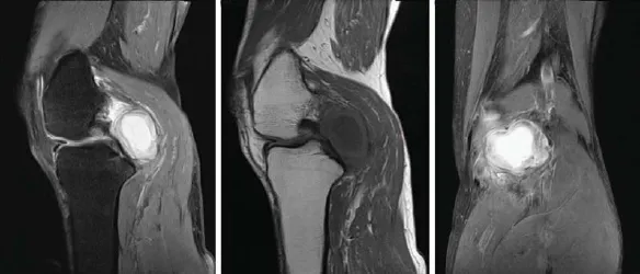
Figure 1. Magnetic resonance images of a 55-year-old female with solitary extraskeletal chondroma (arrows) in the left popliteal region.
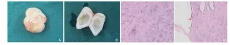
Figure 2. The gross view showed that the size of the tumor was about 4.0 cm × 3.7 cm × 3.0 cm (A). The cut surface showed the cyst cavity in the center of the mass was filled with mucus (B). HE staining revealed the tumor tissue was composed of mature hyaline cartilage and fibrocartilage, with unobvious calcification and different levels of mucoid and cystic degeneration. Chondroma cells exhibited polymorphic without nuclear atypia and mitosis (C); mucoid degeneration was observed in the center of the tumor (arrow, D). ×100
REFERENCES
1. Ishii T, Ikeda M, Oka Y. Subungual extraskeletal chondroma with finger nail deformity: case report. J Hand Surg Am 2010; 35:296-9.
2. Zlatkin MB, Lander PH, Begin LR, et al. Soft-tissue chondromas. AJR Am J Roentgenol 1985; 144:1263-7.
3. Nehete R, Nehete A, Singla S, et al. Soft tissue chondroma of hard palate associated with cleft palate. Indian J Plast Surg 2012; 45:550-2.
4. Aslam MB, Haqqani MT. Extraskeletal chondroma of parotid gland. Histopathology 2006; 48:465-7.
5. Smida M, Abdenaji W, Douira-Khomsi W, et al. Childhood soft tissue chondroma. Two cases report. Tunis Med 2011; 89:379-82.
6. Aslam MB, Haqqani MT. Extraskeletal chondroma of parotid gland. Histopathology 2006; 48:465-7.
7. Kostopoulos IS, Daniilidis I, Velegrakis G, et al. Chondroma of the parotid gland. Clinical histologic immunohistochemical findings of a rare case. Laryngorhinootologie 1993; 72:261-3.
for publication March 15, 2015.
Tel: 86-13762696791, E-mail: kaoyan1982@ yeah.net
 Chinese Medical Sciences Journal2015年4期
- Chinese Medical Sciences Journal的其它文章
- Pseudophakic Malignant Glaucoma Treatment Assisted with Anterior Segment Optical Coherence Tomography: A Case Report
- Inhibition Mechanism of Novel Pyrazolo[1,5-a]pyrazin-4(5H)-one Derivatives Against Proliferation of A549 and H322 Cancer Cells△
- Influence of Photodynamic Therapy on Apoptosis and Invasion of Human Cholangiocarcinoma QBC939 Cell Line
- Effect of Atorvastatin on Expression of Peroxisome Proliferator-activated Receptor Beta/delta in AngiotensinⅡ-induced Hypertrophic Myocardial Cells In Vitro△
- Retroperitoneal Versus Transperitoneal Laparoscopic Partial Nephrectomy: A Systematic Review and Meta-analysis△
- Gender Differences in Ventricular-vascular Coupling Following Exercise△