
Polyketide and benzopyran compounds of an endophytic fungus isolated from Cinnamomum mollissimum: biological activity and structure
2014-03-22 13:01:30CarolinaSantiagoLinSunMurrayHerbertGibsonMunroJacintaSanthanam
Carolina Santiago, Lin Sun, Murray Herbert Gibson Munro, Jacinta Santhanam*
1Biomedical Science Programme, School of Diagnostic and Applied Health Sciences, Faculty of Health Sciences, Universiti Kebangsaan Malaysia, Jalan Raja Muda Abdul Aziz, 50300 Kuala Lumpur, Malaysia
2Department of Chemistry, University of Canterbury, Private Bag 4800, Christchurch 8140, New Zealand
Polyketide and benzopyran compounds of an endophytic fungus isolated from Cinnamomum mollissimum: biological activity and structure
Carolina Santiago1, Lin Sun2, Murray Herbert Gibson Munro2, Jacinta Santhanam1*
1Biomedical Science Programme, School of Diagnostic and Applied Health Sciences, Faculty of Health Sciences, Universiti Kebangsaan Malaysia, Jalan Raja Muda Abdul Aziz, 50300 Kuala Lumpur, Malaysia
2Department of Chemistry, University of Canterbury, Private Bag 4800, Christchurch 8140, New Zealand
PEER REVIEW
Peer reviewer
Siti Alwani Ariffin, Faculty of Pharmacy, University Teknologi MARA (UiTM), 42300 Bandar Puncak Alam, Selangor Darul Ehsan, Malaysia.
E-mail: ctalwani@gmail.com
Comments
This study has studied the bioactivity of three compounds isolated from Phoma sp. fungus. The findings are well represented which indicated endophytic fungi may have a significance meaning in the drug discover.
Details on Page 631
Objective:To study bioactivity and compounds produced by an endophytic Phoma sp. fungus isolated from the medicinal plant Cinnamomum mollissimum.
Endophytic fungi, HPLC bioactivity profiling, Cinnamomum mollissimum, Antimicrobial
1. Introduction
In 1996, the discovery of taxol, an anticancer compound (originally isolated from the plantTaxus brevifolia) from an endophytic fungal speciesPestalotiopsis microspora, highlighted endophytic fungi as an alternate source for drug discovery[1]. Following this discovery, many studies reported the enormous potential of natural products from endophytes which exhibited antimicrobial, antiviral, antiparasitic and antitumor properties[2]. Endophytic fungi are described as fungi which live within plant tissue with no evidence of symptoms to the host plant. Some of these endophytes provide protection to their host plant from tissue invading pathogens by producing compounds that are involved thehost plant’s defense mechanism[3-5]. The fungi have been shown to produce similar compounds as their host plant[6,7]. This provided the rationale to investigate the bioactivity of compounds produced by endophytes, especially those isolated from medicinal plants.
Plants ofCinnamomumspecies (sp.) are known to posses antimicrobial activities and are widely applied in herbal therapy in treating colds, sinusitis, bronchitis and fungal infection[8,9]. A wide range of biological activities ranging from antiseptic, antitumor to antifungal were reported to be found in constituents and essential oils ofCinnamomumsp. plants[10-12]. In this research, an endophytic fungus isolated fromCinnamomum mollissimum(C. mollissimum) was studied. The fungal isolate CB 007 [water agar (WA)], aPhomasp., was previously investigated, and found to produce 5-hydroxyramulosin, a compound that was active againstAspergillus niger(A. niger) (IC501.56 μg/mL) and cytotoxic against murine leukemia cells (IC502.10 μg/mL)[13]. In this study, we report the isolation and biological activity of other compounds produced by CB 007 (WA).
2. Materials and methods
2.1. Sampling of C. mollissimum
Plant parts ofC. mollissimumwere obtained from Universiti Kebangsaan Malaysia Forest Reserve, in Bangi, Selangor, Malaysia. Several twig samples were sampled from a young tree approximately 1.3 m tall. Samples were stored at 4 °C until further processing. The plant specimen voucher (No. 955) was deposited in the herbarium of Universiti Kebangsaan Malaysia and identified by university’s botanist.
2.2. Isolation of endophytes
Plant samples were processed according to methods described by Jayanthiet al. 2011[14] and incubated on potato dextrose agar and WA at 27 °C. Emergence of hyphal tips was periodically checked for 3-14 d. The individual hypal tips were removed and cultured on potato dextrose agar at 27 °C and culture purity was monitored regularly. One of the fungal isolates, CB 007 (WA) was selected for further experimentation, based on the preliminary screening results[13].
2.3. Fermentation and extraction of fungal metabolites
The fungal isolate was cultured in 1 000 mL Erlenmeyer flask containing 200 mL of potato dextrose broth, shaken at 140 r/min, 27 °C for 14 d. Mycelia were removed via filtration and the culture broth was extracted with an equal amount of ethyl acetate overnight. The filtrate was concentrated by removing the solvents under reduced pressure at 35-40 °C with a rotary evaporator (Büchi, New Castle, USA). The concentrated crude extract was ensured to be dry and stored at 4 °C.
2.4. High performance liquid chromatography (HPLC) analysis
An aliquot of the crude extract (250 μg) was analyzed by reverse phase C18HPLC using a gradient solvent system comprising acetonitrile and ultra-pure water as described in methods by Santiagoet al.2012[13]. HPLC was performed on a Dionex (Sunnyvale, USA) system equipped with an ISCO Foxy Jr. sample collector using a reversed-phase analytical column (Phenomenex Prodigy C18, 4.6 mm×250 mm, 5 μm) with photodiode array and an Alltech evaporative light scattering detection (ELSD) (Grace, Deerfield, USA).
2.5. Bioactivity profiling-cytotoxicity assay
Fractions from crude extract (88 fractions×200 μL) were collected in a microtiter plate from HPLC analysis. Daughter plates were made by transferring 50 μL from the original microtiter plate to another plate for analysis of cytotoxicity against P388 murine leukemic cells. The solvent was evaporated prior to the assay by using a centrifugal evaporator. Medium used for the cytotoxicity assay was β-methoxyethoxymethyl, fetal calf serum (10%), penicillin (266 μg/mL), streptomycin (132 μg/mL), L-glutamine (0.002 mol/L), sodium bicarbonate (2.2 g/L), and 4-(2-hydroxyethyl)-1-piperazineethanesulfonic acid (0.0074 mol/L). The plate was incubated 36 °C for 3 d. To obtain the result, 20 μL of thiazolyl blue tetrazolium solution (3.8 mg/mL in phosphate buffered saline) was added to every well and the plate was incubated for 4 h at 36 °C. After incubation, hydrochloric acid in isopropanol (170 μL, 0.08 mol/L) was used to dissolve the formazon product. Cell viability was determined by measuring the absorbance of every well at 540 nm. The absorbance of cell free control and the analyte free cell control was taken as 0% and 100% growth reference, respectively. Cisplatin was used as reference positive control in this experiment.
2.6. Bioactivity profiling-antimicrobial assay
The assay was done according to methods described in Santiagoet al. 2012[13]. A second microtiter plate containing fractions (88 fractions×200 μL) was obtained from HPLC analysis and assayed directly after evaporating the solvent with a centrifugal evaporator. Each well was added with 10 μL of 5% v/v methanol in water and 50 μL growth media. Media used for antifungal assay was RPMI-1640 and for antibacterial assay was Mueller Hinton broth. This was followed by addition of 40 μL of suspension of test organisms. The inoculum size of test organisms used was 2.5×10³CFU/mL for the pathogenic fungiA. nigerandA.fumigatusand 5×10⁵CFU/mL forBacillus subtilis(B. subtilis) bacteria. Plates were incubated at 27 °C, 48 h forA. nigerandA. fumigatus; 37 °C, 24 h forB. subtilis. After incubation, an tetrazolium solution (20 μL, 5 mg/mL) was added to all wells and plates were further incubated (27 °C, 4 h). Then, 100 μL of dimethyl sulfoxide was added into every well to dissolve the formazon product. Cell viability percentage was determined by measuring the absorbance of every well at 540 nm and subtracting the absorbance of cell free control. The absorbance of cell free control (solvent solution+medium) and the analyte free cell control (solvent solution+test organisms in medium) was taken as 0% and 100% growth reference, respectively. Cell growth inhibition was calculated as 100%-cell viability%. The antibiotic gentamycin and antifungal amphotericin B used as positive controls in this experiment.
2.7. Isolation of compounds
The crude extract was subjected to HPLC by injecting 250 μg of extract onto a C18column (250 mm×4.6 mm, 5 μm, Phenomenex Luna) using the same gradient solvent system as before for HPLC analysis. The collection of the bioactive fractions was scaled up to 10 times, to collect the pure compounds.
2.8. Determination of compounds
The pure compounds were subjected to nuclear magnetic resonance (NMR) and mass spectrometry experiments were for identification. The NMR experiments was recorded on a Varian INOVA 500 spectrometer (Varian, Palo Alto, USA) at 23 °C, operating at 500 MHz at 23 °C, using a capillary probe. High resolution electron impact mass spectra were obtained on a liquid chromatography time-time of flight mass spectrometer (Micromass, Greater Manchester, UK). Structural elucidation of compounds process was aided by access to AntiMarin database.
3. Results
In bioactivity profiling, HPLC fractions were assayed on a microtiter plate for cytotoxicity and antimicrobial activity. A line graph was plotted to quantitatively indicate the inhibitory activity of HPLC fractions. These fractions correspond directly to collection time over 40 min from fraction 1 being the first collected fraction to fraction 88 the last collected fraction. Figure 1 is showing bioactivity profile for first 36 HPLC fractions obtained from CB 007 (WA). The remaining 46 fractions which eluted later (after fraction 36) did not exhibit any biological activity; hence the results were not included in Figure 1. An active region shown on the graph was interpreted as where most of the biological activity was found. The active region for CB 007 (WA) extract was from fraction 28 to 34 in which it was observed inhibitory activities against P388 murine leukemic cells,B. subtilisandA. niger(Figure 1). Several few fractions which eluted earlier (fraction 3 to 13) indicated the potent antifungal activity againstA. fumigatus.
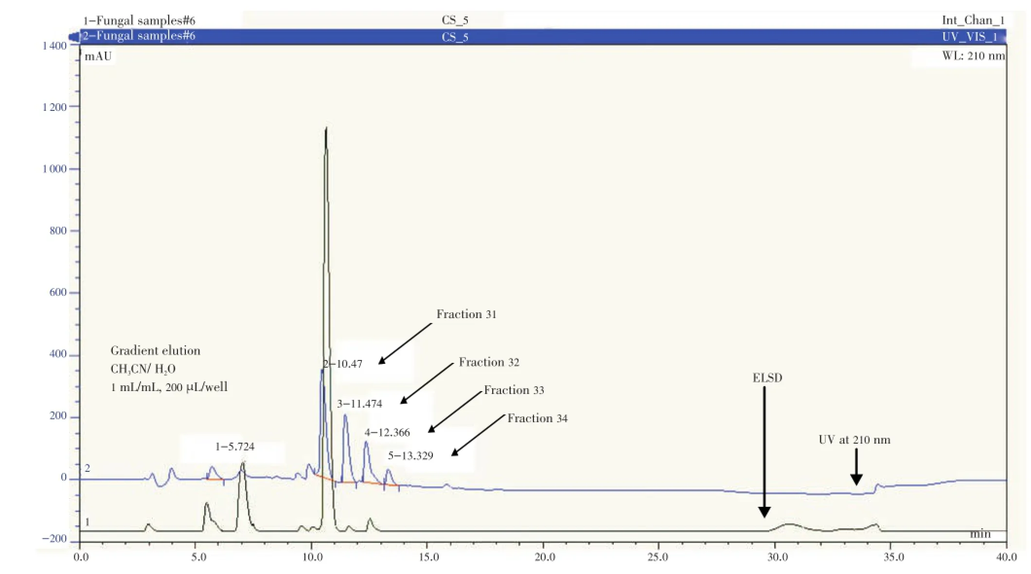
Figure 2. HPLC chromatogram with ELSD and UV detection, of 250 μg fungal endophyte CB 007 (WA) extract, showing major chromatograph peaks present in the biologically active region consisting fraction 31, 32, 33 and 34 which correlated to collection time between 10 to 14 min.
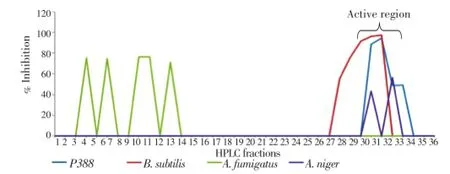
Figure 1. Bioactivity profile of HPLC fractions (fraction 1 to 36) from the fungal endophyte CB 007(WA) extract showing inhibition of P388 murine leukemic cells, B. subtilis, and A. niger in active region.An active region is interpreted as region with most bioactivity observed as indicated by the marked area in the graph.
The bioactivity profiling led to identification of active region which directly corresponded to a specific time region between 10 to 14 min in the HPLC chromatogram (Figure 2). Four major peaks were present in the chromatogram in this time frame and each fraction that correlated to the peaks were collected and purified. This led to isolation of 4-hydroxymellein (A), 4,8-dihydroxy-6-methoxy-3-methyl-3,4-dihydro-1H-isochromen-1-one (B) and 1-(2,6-dihydroxyphenyl) ethanone (C) as depicted in Figure 3. The compound A, B and C eluted after 11.5 (fraction 32), 12.4 (fraction 33) and 13.2 (fraction 34) min, respectively (Figure 2). The compound 5-hyroxyramulosin has been reported previously[13,15] eluted after 10.47 min (fraction 31). The spectral data for compounds A, B and C is as following:
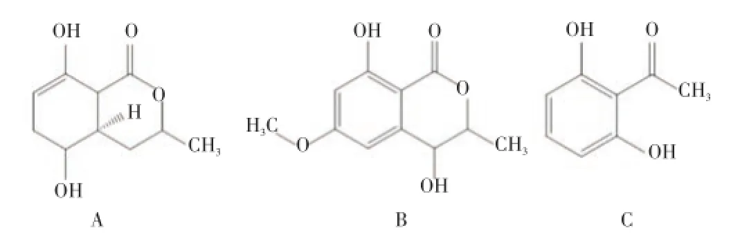
Figure 3. Structure of 4-hydroxymellein (A), 4,8-dihydroxy-6-methoxy-3-methyl-3,4-dihydro-1H-isochromen-1-one (B) and 1-(2,6-dihydroxyphenyl) ethanone (C).
4-Hydroxymellein (A): white solids; UV (MeOH) λmax210, 245, 314 nm. HRESIMSm/z193.0685 [M+H]+.1H and13C NMR data were consistent to previously reported literature[16,17]; Formula C10H10O4.
4,8-Dihydroxy-6-methoxy-3-methyl-3,4-dihydro-1H-isochromen-1-one (B): white solids; UV (MeOH) λmax215, 266, 301 nm. HRESIMSm/z255.0744 [M+H]+. Natural compound for stock screening; Formula C11H12O5.
1-(2,6-dihydroxyphenyl) ethanone (C): white solids; UV (MeOH) λmax210, 243, 310 nm. HRESIMSm/z142.0443 [M].1H and13C NMR data were consistent to previously reported literature[18]; Formula C8H8O3.
The biological activity of bioactive HPLC fractions from CB 007 (WA) and compounds isolated from them is summarized in Table 1. Compound A has inhibitory activity against P388 murine leukemic cells andB. subtilis. Compound B was inhibiting P388 murine leukemic cells andA. niger. However, compound C was inactive in all tested assays.
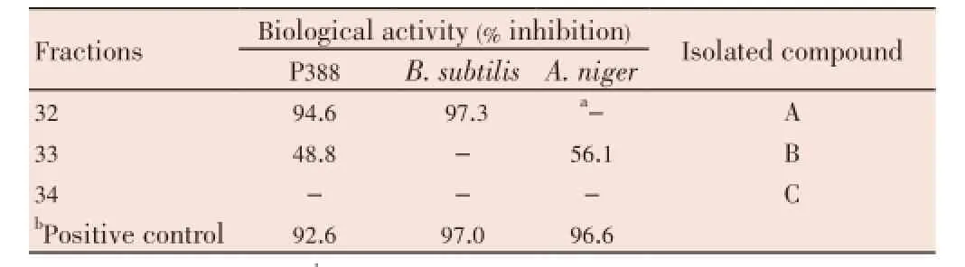
Table 1 Biological activity and major compounds isolated from HPLC fractions of the fungal endophyte CB007 (WA) extract.
4. Discussion
Endophytes are known to produce pharmacologically active compounds. The strategy in this research was to explore endophytic fungi from a medicinal plant.Cinnamomumplants are widely utilized as natural remedies to cure many diseases because of their healing properties. In herbal therapeutics, essential oils from the leaf, inner bark and stems of the plant are used as remedies[10]. Biologically active compounds from endophytes ofCinnamomumsp. have been studied previously but there is no reported study to date on endophytes fromC. mollissimum.
The endophyte CB 007 (WA) showed encouraging antimicrobial and cytotoxic activity in a screening assay[13]. Therefore, the research was undertaken to identify the compounds that gave rise to the observed activity. Metabolites from the isolate were extracted and analysed. This eventually led to the isolation of three known compounds; 4-hydroxymellein, 4,8-dihydroxy-6-methoxy-3-methyl-3,4-dihydro-1H-isochromen-1-one and 1-(2,6-dihydroxyphenyl) ethanone.
The compounds 4-hydroxymellein and 1-(2,6-dihydroxyphenyl) ethanone are polyketides that are synthesized by the polyketide synthase pathways. 4-hydroxymellein was first isolated from a fungus,Cercospora taiwanesis. Fungus belonging to this genus is known to be pathogenic to crops such as sugarbeets and soyabeans[16]. A recent research also reported the isolation of 4-hydroxymellein from an endophytic fungus ofPenicilliumsp., which exhibited active anti-fungal activities[19]. Meanwhile, the compound 1-(2,6-dihydroxyphenyl) ethanone was firstly discovered fromDaldinia concentrica, a fungus belonging to Ascomycota division[18]. No biological activities has been reported for this compound. Polyketides compounds such as the above mentioned compounds are results of condensation of activated primary metabolites (acetyl-CoA and malonyl-CoA) to form b-ketoacetyl polymers which are linked to the enzyme by thioester bonds. They are structurally and functionally related to fatty acid synthases[20] and are identified by a common biosynthetic origin of carbon atoms derived from small carboxylicacids[21]. Polyketides are frequently found as fungal metabolites and they are an important class of compounds in natural product drug discovery due to their structural diversity. They possess remarkable biologically activity as antibiotic, antifungal, anticancer, antiparasitic, and immunosuppressant[22]. Examples of polyketides that have been commercialized include amphotericin B (antifungal), lovastatin (anti-cholesterol), rapamycin (immunosuppressant) and erythromycin B (antibiotic). As such, the two polyketides compounds A and C can be potentially developed into drug in medicine industry.
Compounds of mellein derivatives have been reported to display antimicrobial and cytotoxic activity which is supported by the observation in this study[23]. 4-hydroxymellein demonstrated inhibitory activity againstB. subtilisand P388 murine leukemic cells (97.3% and 94.6% respectively) which has not been reported before. However, 1-(2,6-dihydroxyphenyl) ethanone was inactive for the tested activity. Assays conducted in this research were limited to evaluate antibacterial, antifungal and leukemia cytotoxic activity. As such, otherin vitroassays such as antitumor or anti-parasitic assays can be employed to fully examine the potential of 1-(2,6-dihydroxyphenyl) ethanone.
The compound 4,8-dihydroxy-6-methoxy-3-methyl-3,4-dihydro-1H-isochromen-1-one showed moderate inhibitory activity against P388 murine leukemic cells (48.8%) andA. niger(56.1%) and no other biological activities were reported for this particular compound found from the literature review. This compound belongs to the benzopyran group which is an organic compound that is produced by fusion of benzene ring to heterocyclic pyran ring. Since benzopyrans are associated with polyketide synthesis this compound may have arisen from the same carbon framework as 4-hydroxymellein and 1-(2,6-dihydroxyphenyl) ethanone via polyketide synthase action. Among natural products, benzopyrans were found to be the core ring structures of flavonoids, isoflavanoids and isocoumarins[24]. Benzopyran derivatives possess numerous pharmocological properties as diuretic, analgesic, myorelaxant, and hypoglycaemic[25-27]. Besides this, benzopyrans are potential intermediates in the synthesis of steroid analogs, or function as building blocks in the production of pterocarpans and isoflavones that have strong fungicidal activity[28]. Hence, the fungicidal activity of compound B observed in this study is attributed to its structural and functional similarities to pterocarpans and isoflavones.
Although, this study reports three known compounds, new biological activities have been identified in these compounds. In addition, the activity of the compounds is specific for either bacteria or fungi alone unlike toxic compounds which destroy all cell types. Therefore, further studies are needed to exploit the full potential of these compounds as antifungal or antibacterial agent. We conclude that endophytic fungi from medicinal plants is a significant resource for drug discovery.
Conflict of interest statement
We declare that we have no conflict of interest.
Acknowledgements
Financial support for this study from Universiti Kebangsaan Malaysia, Research University Grant UKMGUP-SK-08-23-300, and Science Fund 02-01-02-SF 0517 is acknowledged. We thank Department of Chemistry, University of Canterbury and Department of Pharmacology, Universiti Malaya for technical assistance provided in chemistry experiments.
Comments
Background
The present investigation demonstrates the bioactivity of the three known compounds isolated fromPhomasp. inC. mollissimum. This may lead to further research and explore on the potential of endopytic fungal (Phomasp.) and their active compounds.
Research frontiers
The author found three actives compounds fromPhomasp. inC. mollissimumand its biological activity.Phomasp. extract contained 4-Hydroxymellein, 4,8-Dihydroxy-6-methoxyl-3,4-dihydro-1H-isochromen-1-one and 1-(2,6-dihydroxyphenyl) ethanone. These compounds exihibited inhibition against P388,B. subtilisandA. niger.
Related reports
So far, many research have been done on the isolation of endophytic fungi from medicinal plants. The present work revealed on the three known compounds isolated fromPhomasp. with theirs bioactivity.
Innovations and breakthroughs
Several studies were carried out on the isolation of endophytic fungi fromC. mollissimum. In the present study, authors have worked on the isolation of three active compounds fromPhomasp. endophytic fungi.
Applications
The present investigation demonstrates the bioactivity of the three known compounds isolated fromPhomasp. inC. mollissimum. This may lead to further research and explore on the potential of endopytic fungal (Phomasp.) and their active compounds.
Peer review
This study has studied the bioactivity of three compoundsisolated fromPhomasp. fungi. The findings are well represented which indicated endophytic fungi may have a significance meaning in the drug discover.
[1] Amal HA, Abdessamad D, Kjer J, Proksch P. Fungal endophytes from higher plants: a prolific source of phytochemicals and other bioactive natural products. Fungal Divers 2010; 41(1): 1-16.
[2] Kusari S, Hertweck C, Spiteller M. Chemical ecology of endophytic fungi: origins of secondary metabolites. Chem Biol 2012; 19(7): 792-798.
[3] Hyde KD, Soytong K. The fungal endophyte dilemma. Fungal Divers 2008; 33: 163-173.
[4] Purahong W, Hyde KD. Effects of fungal endophytes on grass and non-grass litter decomposition rates. Fungal Divers 2011; 47(1): 1-7.
[5] Krings M, Taylor TN, Dotzler N. Fungal endophytes as a driving force in land plant evolution: evidence from the fossil record. In: Southworth D, editor. Biocomplexity of plant-fungal interactions. New Jersey: John Wiley & Sons Inc; 2012, p. 1-28.
[6] Zhao J, Shan T, Mou Y, Zhou L. Plant-derived bioactive compounds produced by endophytic fungi. Mini-Rev Med Chem 2011; 11(2): 159-168.
[7] Strobel G. Genetic diversity of microbial endophytes and their biotechnical applications. In: Nelson KE, Jones-Nelson B, editors. Genomics applications for the developing world. New York: Springer; 2012, p. 249-262.
[8] Kharwar RN, Maurya AL, Verma VC, Kumar A, Gond SK, Mishra A. Diversity and antimicrobial activity of endophytic fungal community isolated from medicinal plant Cinnamomum camphora. Proc Natl Acad Sci India Sect B Biol Sci 2012; 82(4): 557-565.
[9] Rahmatullah M, Khatun A, Morshed N, Neogi PK, Khan SUA. A randomized survey of medicinal plants used by folk medicinal healers of Sylhet Division, Bangladesh. Adv Nat Appl Sci 2010; 4: 52-62.
[10] Unlu M, Ergene E, Unlu GV, Zeytinoglu HS, Vural N. Composition, antimicrobial activity and in vitro cytotoxicity of essential oil from Cinnamomum zeylanicum Blume (Lauraceae). Food Chem Toxicol 2010; 48(11): 3274-3280.
[11] Chen CY, Chen CH, Lo YC, Wu BN, Wang HM, Lo WL, et al. Anticancer activity of isoobtusilactone a from Cinnamomum kotoense: involvement of apoptosis, cell-cycle dysregulation, mitochondria regulation, and reactive oxygen species. J Nat Prod 2008; 71(6): 933-940.
[12] Jantan I, Karim M, Santhanam J, Jamal JA. Correlation between chemical composition and antifungal activity of the essential oils of eight Cinnamomum species. Pharmcol Biol 2008; 46(6): 406-412.
[13] Santiago C, Fitchett C, Munro MHG, Jalil J, Santhanam J. Cytotoxic and antifungal activities of 5-hydroxyramulosin, a compound produced by an endophytic fungus isolated from Cinnamomum mollisimum. Evid Based Compl Alt Med 2012; 2012: 1-6.
[14] Jayanthi G, Kamalraj S, Karthikeyan K, Muthumary J. Antimicrobial and anti antioxidant activity of the endophytic fungus Phomopsis sp. GJJM07 isolated from Mesua ferrea. Int J Curr Res 2011; 1: 85-90.
[15] Osterhage C, K?nig GM, Jones PG, Wright AD. 5-hydroxyramulosin, a new natural product produced by Phoma tropica, a marinederived fungus isolated from the alga Fucus spiralis. Planta Med 2002; 68(11): 1052-1054.
[16] Camarda L, Merlini L, Nasini G. Metabolites of Cercospora. Taiwapyrone, an α-pyrone of unusual structure from Cercospora taiwanensis. Phytochemistry 1976; 15(4): 537-579.
[17] Assante G, Locci R, Camarda L, Merlini L, Nasini G. Screening of the genus Cercospora for secondary metabolites. Phytochemistry 1977; 16(2): 243-247.
[18] Allport DC, Bu’Lock JD. Biosynthetic pathways in Daldinia concentrica. J Chem Soc 1960; 134: 654-662.
[19] Oliveira CM, Silva GH, Regasini LO, Zanardi LM, Evangelista AH, Young MC, et al. Bioactive metabolites produced by Penicillium sp. 1 and sp. 2, two endophytes associated with Alibertia macrophylla (Rubiaceae). Z Naturforsch C 2009; 64(11-12): 824-830.
[20] Miller KI, Qing C, Sze DM, Roufogalis BD, Neilan BA. Culturable endophytes of medicinal plants and the genetic basis for their bioactivity. Microb Ecol 2012; 64(2): 431-449.
[21] Yu D, Zeng J, Chen D, Zhan J. Characterization and reconstitution of a new fungal type III polyketide synthase from Aspergillus oryzae. Enzyme Microb Tech 2010; 46(7): 575-580.
[22] Debbab A, Aly AH, Proksch P. Bioactive secondary metabolites from endophytes and associated marine derived fungi. Fungal Divers 2011; 49(1): 1-12.
[23] Klaiklay S, Rukachaisirikul V, Sukpondma Y, Phongpaichit S, Buatong J, Bussaban B. Metabolites from the mangrove-derived fungus Xylaria cubensis PSU-MA34. Arch Pharmacal Res 2012; 35(7): 1127-1131.
[24] Cankarova N, Krchnak V. Solid-phase synthesis enabling chemical diversity. In: Trabocchi A, editor. Diversity-oriented synthesis: basics and applications in organic synthesis, drug discovery, chemical biology. New Jersey: John Wiley & Sons Inc; 2013, p. 201-252.
[25] Ghorbani-Vaghei R, Toghraei-Semiromi Z, Karimi-Nami R. One-pot synthesis of 4H-Chromene and Dihydropyrano [3,2-c] chromene derivatives in hydroalcoholic media. J Braz Chem Soc 2011; 22(5): 905-909.
[26] Thomas N, Zachariah SM. Pharmacological activities of Chromene derivatives: an overview. Asian J Pharm Clin Res 2013; 6(2): 11-15.
[27] Tang L, Yang YS, Ji RY. Synthesis and biological activity of a series of benzopyran derivatives. Yao Xue Xue Bao 2008; 43(2): 162-168.
[28] Tripathi AK, Mukherjee D, Koul S, Taneja SC, Agrawal SK, Sharma PR, et al. Synthesis and cytotoxity of novel benzopyran derivatives. Indian J Chem Sec B 2011; 50(11): 1619-1629.
10.12980/APJTB.4.2014APJTB-2014-0030
*Corresponding author: Jacinta Santhanam, Biomedical Science Programme, School of Diagnostic & Applied Health Sciences, Faculty of Health Sciences, Universiti Kebangsaan Malaysia, Jalan Raja Muda Abdul Aziz, 50300 Kuala Lumpur, Malaysia.
Tel: +603-92897039
Fax: +603-26929032
E-mail: jacinta@medic.ukm.my
Foundation Project: Supportted by Universiti Kebangsaan Malaysia, Research University Grant UKM-GUP-SK-08-23-300, and Science Fund 02-01-02-SF 0517.
Article history:
Received 22 May 2014
Received in revised form 8 Jun, 2nd revised form 15 Jun, 3rd revised form 23 Jun 2014
Accepted 22 Jul 2014
Available online 28 Aug 2014
Methods:Compounds produced by the fungus were extracted from fungal broth culture with ethyl acetate. This was followed by bioactivity profiling of the crude extract fractions obtained via high performance liquid chromatography. The fractions were tested for cytotoxicity to P388 murine leukemic cells and antimicrobial activity against bacteria and pathogenic fungi. Compounds purified from active fractions which showed antibacterial, antifungal and cytotoxic activities were identified using capillary nuclear magnetic resonance analysis, mass spectrometry and admission to AntiMarin database.
Results:Three known compounds, namely 4-hydroxymellein, 4,8-dihydroxy-6-methoxy-3-methyl-3,4-dihydro-1H-isochromen-1-one and 1-(2,6-dihydroxyphenyl) ethanone, were isolated from the fungus. The polyketide compound 4-hydroxymellein showed high inhibitory activity against P388 murine leukemic cells (94.6%) and the bacteria Bacillus subtilis (97.3%). Meanwhile, 4,8-dihydroxy-6-methoxy-3-methyl-3,4-dihydro-1H-isochromen-1-one, a benzopyran compound, demonstrated moderate inhibitory activity against P388 murine leukemic cells (48.8%) and the fungus Aspergillus niger (56.1%). The second polyketide compound, 1 (2,6-dihydroxyphenyl) ethanone was inactive against the tested targets.
Conclusions:These findings demonstrate the potential of endophytes as producers of pharmacologically important compounds, including polyketides which are major secondary metabolites in fungi.
 Asian Pacific Journal of Tropical Biomedicine2014年8期
- Asian Pacific Journal of Tropical Biomedicine的其它文章
- Adult Klebsiella pneumoniae meningitis in Qatar: clinical pattern of ten cases
- Iron-chelating and anti-lipid peroxidation properties of 1-(N-acetyl-6-aminohexyl)-3-hydroxy-2-methylpyridin-4-one (CM1) in longterm iron loading β-thalassemic mice
- Digestive fungal flora in asymptomatic subjects in Bobo-Dioulasso, Burkina Faso
- Glucose-6-phosphate dehydrogenase (G6PD) deficiency is associated with asymptomatic malaria in a rural community in Burkina Faso
- GC/GCMS analysis of the petroleum ether and dichloromethane extracts of Moringa oleifera roots
- An efficient method in breaking of dormancy from Bunium persicum (Boiss) Fedtsch seeds: a valuable herb of Middle East and Central Asia