
Histopathological and molecular study of Neospora caninum infection in bovine aborted fetuses
2014-03-21 07:49:50AmirKamaliHesamAdinSeifiAhmadRezaMovassaghiGholamRezaRazmiZahraNaseri
Amir Kamali, Hesam Adin Seifi,*, Ahmad Reza Movassaghi, Gholam Reza Razmi, Zahra Naseri
1Department of Clinical Sciences, Faculty of Veterinary Medicine, Ferdowsi University of Mashhad, Mashhad, Iran
2Center of Excellence in Ruminant Abortion and Neonatal Mortality, Ferdowsi University of Mashhad, Mashhad, Iran
3Department of Pathobiology, Faculty of Veterinary Medicine, Ferdowsi University of Mashhad, Mashhad, Iran
Histopathological and molecular study of Neospora caninum infection in bovine aborted fetuses
Amir Kamali1, Hesam Adin Seifi1,2*, Ahmad Reza Movassaghi2,3, Gholam Reza Razmi2,3, Zahra Naseri2
1Department of Clinical Sciences, Faculty of Veterinary Medicine, Ferdowsi University of Mashhad, Mashhad, Iran
2Center of Excellence in Ruminant Abortion and Neonatal Mortality, Ferdowsi University of Mashhad, Mashhad, Iran
3Department of Pathobiology, Faculty of Veterinary Medicine, Ferdowsi University of Mashhad, Mashhad, Iran
ARTICLE INFO
Article history:
Received 9 Sep 2014
Received in revised form 23 Sep 2014
Accepted 7 Oct 2014
Available online 17 Oct 2014
Neospora caninum
Dairy herds
Abortion
PCR
Histopathology
Risk factors
Objective:To estimate the extent to which abortion in dairy cows was associated with ofNeospora caninum(N. caninum) and to determine the risk factors of neosporosis in dairy farms from 9 provinces in Iran.
1. Introduction
Neospora caninum(N. caninum), an apicomplexan protozoan, is an important factor of fetal abortion in cattle that causes economic losses in dairy herds worldwide[1]. For the first time in 1984, this parasite was described in puppies with encephalomyelitis and myositis[2]. Historically in ruminant, the first report ofN. caninuminfection was diagnosed in a congenitally infected lamb in England[3]. The main route of transmission in cattle is vertical (80%-100%), although cows could be infected by ingestion of oocysts excreted by dogs and abortion may be observed in any age of pregnancy[4,5]. Dogs are final and intermediate host forN. caninum[6]. It seems that the abortion rate in infected animals is three to seven times higher compared to uninfected ones. The main sign ofNeosporainfection in adult cow is abortion and it most occurs at 5-6 months gestation.N. caninumcan also cause repeated abortion in cows[1]. Symptoms have been reported in calves younger than 2 months. These symptoms include neurological symptoms, weight loss and ataxia. In examination of the nervous system, decreased knee reflex and decreased peripheral sensation is evident. Calves may have protruding or asymmetric eyes.N. caninumoccasionally cause birth defects such as hydrocephalus, spinal canal narrowing and scoliosis[1].N. caninumDNA has been detected by polymerase chain reaction (PCR) and immunohistochemistry methods in brains of aborted fetusesin Iran[4,7]. Seroepidemiological studies have shown that the prevalence ofNeosporainfection is relatively high in dairy cattle[8-10] and dogs in Iran[11,12].The objectives of the presentstudy were to estimate the extent to which abortion in dairy cows was associated with ofN. caninumand to determine the risk factors of neosporosis in dairy farms in Iran. The present study is the first comprehensive assessment of abortion associated withN. caninumin dairy herds in Iran.
2. Materials and methods
The study was performed in 9 provinces (including Khorasan Razavi, North Khorasan, South Khorasan, Golestan, Mazandaran, Isfahan, Chaharmahale Bakhtiari, Kermanshah and West Azerbaijan) (Figure 1). The climate of these provinces varies from cold to warm area and they have almost 280 000 cattle in dairy herds. The herd size varied from farm to farm with a range of 20 to 9 000 cattle. Holstein/Friesian was the most common breed of cattle. This study was performed over a 4 -year-period (2009-2013) in 45 dairy herds.
2.1. Sample collection
During 2009 to 2013, 395 aborted bovine fetuses at different stages of gestation were referred to Center of Excellence in Ruminant Abortion and Neonatal Mortality. Firstly, the aborted fetuses were necropsied and samples of brain were collected under aseptic condition. After that, one half of the brain was taken for PCR and another half for histopathology examination. Collected fetal samples were centrifuged at 2 000 ×gfor 10 min and stored at 20 °C freezer until used.
2.2. DNA extraction and PCR
One half of the brain was homogenized with a stirrer, and DNA was extracted from 1 g homogenate sample using the commercial kit (Cinnagen Inc., Iran) according to the manufacturer’s instructions. PCR was performed as described by Mülleret al.[13]. Pair of Np6/Np21 primers (5’ GGG TGT GCG TCC AAT CCT GTA AC 3’ - 5’ CTC GCC AGT CAA CCT ACG TCT TCT 3’) was used to amplify the 337 bp DNA fragment.
2.3. Histopathological examination

Figure 1.Map of Iran-sampling regions.
For the histopathological study, brain tissues of aborted fetuseswere fixed in 10% neutral buffered formalin. Then they were dehydrated through graded alcohols and embedded in paraffin wax. Sections were cut in 5 μm, deparaffinized, rehydrated and stained with hematoxylin and eosin (H & E).Then the stained sections were histopathologically assessed by light microscopy. Brains were examined for determined lesions and evaluated the parasite distribution. The sections were carefully examined at ×100, ×200, ×400 and ×1 000 magnifications, respectively.
2.4. Risk factors and statistical analysis
To identify the risk factors and clarify other factors associated withNeosporainduced abortion in dairy cattle, the risk factors including season, parity of dam, cow’s milk production, history of bovine viral diarrhea (BVD) and infectious bovine rhinotracheitis (IBR) infection in herd, herd size, aborted fetal age, occurrence of stillbirth and fetal appearance were statistically analyzed. Statistical analysis was performed by SAS (Version 9.2).Chisquare analysis and logistic regression model were used to identify risk factors associated withN. caninuminfected abortion.
3. Results
Brain samples of a total 395 aborted fetuses were examined by PCR for detection ofN. caninum. Out of 395 brains samples, 179 samples (45%) were positive (Figure 2).
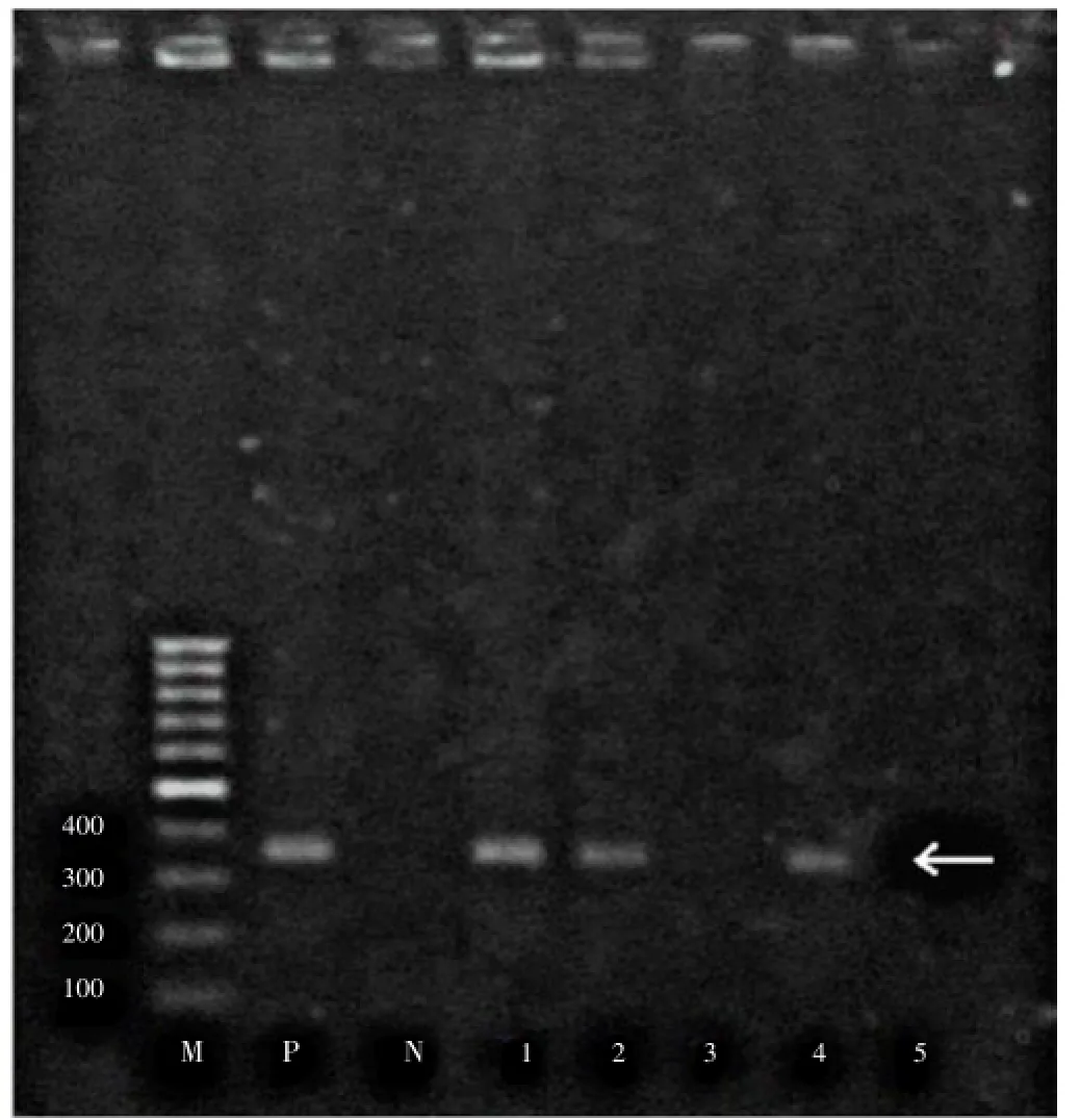
Figure 2.The PCR test for detectingN. caninuminfection.M: Size marker (100 bp DNA Ladder); P: Positive control; N: Negative control; 1-5: Samples; 1, 2, 4:N. caninumpositive; 3, 5:N. caninumnegative. Right arrow shows 337 bp PCR product.
Only 56 fetal brains of PCR-positive were suitable for histopathological examination. Of these 56 samples, 16 (28%) showed the characteristic lesions ofNeosporainfection such as non-suppurative encephalitis. Other patterns of brain damage that observed in this study were non-suppurative meningitis (5%), gliosis (focal and diffuse) (93%), satellitosis (2%), severe hyperemia (100%), hemorrhage (focal and diffuse) (51%), perivascular cuffing (63%), ischemic cell change (25%) and edema (100%). In one case, theNeosporalike cyst with dimensions of 35 μm×47.5 μm was observed (Figure 3).
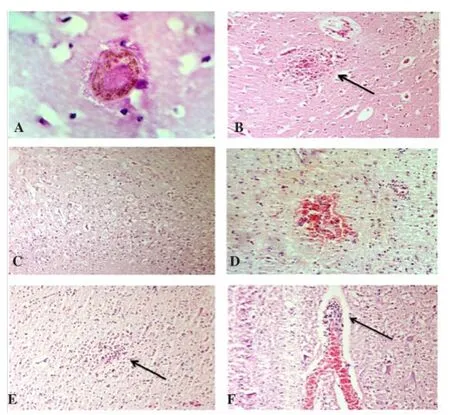
Figure 3.N. caninumlike cyst in the brain.A:N. caninumlike cyst with numerous bradyzoite enclosed in a thick cyst wall in brain (H & E, ×1 000); B: Non-suppurative encephalitis (H & E, ×200); C: Diffuse edema (H & E, ×100); D: Focal hemorrhage in fetus brain parenchyma (H & E, ×200); E: Focal gliosis in fetus brain (H & E, ×200); F: Perivascular cuffing mostly mono nuclear cells in the fetus brain (H & E, ×200).
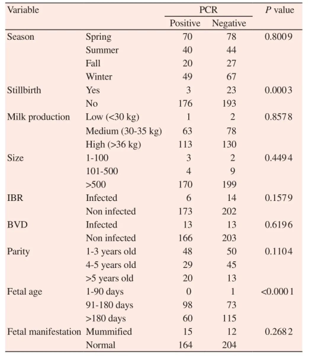
Table 1Chi-sqaure analysis of variables associated toN. caninumabortion based on PCR test.
Statistical analysis indicated that risk factor variables including season, parity of dam, the history of BVD and IBR occurrence in herd, cow’s milk production, herd size and fetal appearance did not show significant association withNeosporacaused abortion based on PCR test (P>0.05), but two variables including fetal age and stillbirth showed a significant association with PCR results (P<0.001) (Table 1).
The stillbirth incidence was significantly less inNeosporainfected fetuses than non-infected ones. In addition, this study showed thatNeosporacaused abortion was significantly more in the second trimester of pregnancy than other periods (P<0.001). All variables were analyzed to the logistic regression model, which indicated that fetal age associated withN. caninum-related abortion was the only significant variable in logistic regression model. The odds of stillbirth occurrence in non-Neosporainfected fetuses were two times greater thanNeosporainfected ones (OR: 2.043, 95%CI: 1.156-3.609,P=0.04).
4. Discussion
In present study, we used PCR and histopathology methods to detectN. caninumin aborted bovine fetuses. Among the diagnostic procedures, PCR was more sensitive and specific than other tests such as histopathology and immunohistochemistry[14] and was less affected by autolysis and postmortem changes[15]. So far, this method has been used for determining the presence ofN. caninumDNA in different embryonic tissues[16], fetal fluids[17,18] and even oocysts in feces of final hosts[19]. PCR results along with histopathological findings in aborted fetal tissues, could confirm the occurrence of abortion byN. caninum. In the European countries and USA, some studies indicated that up to 42% of aborted fetuses from dairy cattle were infected withN. caninum[16]. In some areas of Iran, studies have been done to investigate the contamination rate of the parasite in dairy herds and the infection rate of 18.4%[8], 11.9%[9], and 12.6%[10] were reported. In present study,N. caninumDNA was detected in 45% (179) of brain samples of dairy farms in different provinces of Iran. This rate of infection is higher than other frequencies that were previously reported in Iran.
The main characteristic lesion ofNeosporainfection such as non-suppurative encephalitis[20-22] was observed in 16 (28%) out of 56 positive PCR brain samples. Other lesions in the brain were often focal to diffuse non-suppurative meningitis, mononuclear cell infiltration around acentral area of necrosis, glial proliferation and calcification[16,23]. In present study, other histologic lesions such as non-suppurative meningitis, gliosis, satellitosis, severe hyperemia, hemorrhage, perivascular cuffing, ischemic cell change, and edema were observed in brains of aborted fetuses. In our study, theNeosporalike cyst with dimensions of 35 μm×47.5 μm was observed. In bovine tissues,N. caninumcysts with 5-50 μm in diameter and the thickness of the cyst wall varies from <1 to 2.5 μm was reported[24]. The observed histopathological findings provide a strong evidence forN. caninum-induced abortion in dairy herds of Iran.
Many studies have been done to assess the risk factors associated with neosporosis in dairy cattle[25-29]. We investigated the associations of risk factors including parity, milk production, history of BVD and IBR infection in herd, season, herd size, age of abortions and fetal appearance with abortion due toN. caninumin dairy cows. The results were shown a significant association between fetal age and stillbirth withN. caninuminfection. In the previous studies, there was also a strong relationship between fetal age and stillbirth withNeosporaseroprevalence in dairy
herds[30,31].
According to our results,N. caninumappears to be the most commonly detected and attributable cause of bovine abortion in dairy herds in Iran. Economic losses associated with neosporosis include abortion, neonatal mortality, early fetal death, and the fact that the disease has proved to be challenging to control, add the importance of the neosporosis as a major cause of abortion in dairy farms.
Conflict of interest statement
We declare that we have no conflict of interest.
Acknowledgements
The study was supported by research fund of Ferdowsi University of Mashhad, Mashhad, Iran. (Grant No. 3/25975).All the sampling and molecular diagnoses were performed in Center of Excellence in Ruminant Abortion and Neonatal Mortality. We are very grateful to Mr. Mohhammdnejad for his technical assistance.
[1] Dubey JP, Schares G. Neosporosis in animals-the last five years.Vet Parasiotol2011;180: 90-108.
[2] Salehi N, Haddadzadeh H, Shayan P, Koohi MK. Isolation ofNeospora caninumfrom an aborted fetus of seropositive cattle in Iran.Vet Arhiv2012;82: 545-553.
[3] King JS, Jenkins DJ, Ellis JT, Fleming P, Windsor PA, ?lapeta J. Implications of wild dog ecology on the sylvatic and domestic life cycle ofNeospora caninumin Australia.Vet J2011;188: 24-33.
[4] Razmi GR, Maleki M, Farzaneh N, Talebkhan Garoussi M, Fallah AH. First report ofNeospora caninum-associated bovine abortion in Mashhad Area, Iran.Parasitol Res2007;100: 755-757.
[5] Almería S.Neospora caninumand wildlife.ISRN Parasitol2013; doi: 10.5402/2013/947347.
[6] Monney T, Debache K, Hemphill A. Vaccines against a major cause of abortion in cattle,Neospora caninuminfection.Animals2011;1: 306-325.
[7] Habibi GR, Hashemi-Fesharki R, Sadrebazzaz A, Bozorgi S, Bordbar N. Seminested PCR for diagnosis ofNeospora caninuminfection in cattle.Arch Razi Inst2005;59: 55-64.
[8] Nematollahi A, Moghaddam GH, Jaafari R, Helan JA, Norouzi M. Study on outbreak ofNeospora caninum-associated abortion in dairy cows in Tabriz (Northwest Iran) by serological, molecular and histopathologic methods.Asian Pac J Trop Med2013;6: 942-946.
[9] Razmi GR, Mohammadi GR, Garossi T, Farzaneh N, Fallah AH, Maleki M. Seroepidemiology ofNeospora caninuminfection in dairy cattle herds in Mashhad Area, Iran.Vet Parasitol2006;135: 187-189.
[10] Nourollahi Fard SR, Khalili M, Aminzadeh A. Prevalence of antibodies toNeospora caninumin cattle in Kerman Province, South East Iran.Vet Arhiv2008;78: 253-259.
[11] Haddadzadeh HR, Sadrebazzaz A, Malmasi A, Talei Ardakani H, Khazraii Nia P, Sadreshirazi N. Seroprevalence ofNeospora caninuminfection in dogs from rural and urban environments in Tehran, Iran.Parasitol Res2007;101: 1563-1565.
[12] Hosseininejad M, Hosseini F. Seroprevalence ofNeospora caninumandToxoplasma gondiiinfection in dogs from west and central parts of Iran using two indirect ELISA tests and assessment of associate risk factors.Iran J Vet Res2011;12: 46-51.
[13] Müller N, Zimmermann V, Hentrich B, Gottstein B. Diagnosis ofNeospora caninumandToxoplasma gondiiinfection by PCR and DNA hybridization immunoassay.J Clin Microbiol1996;34: 2850-2852.
[14] Stuart P, Zintl A, De Waal T, Mulcahy G, Hawkins C, Lawton C. Investigating the role of wild carnivores in the epidemiology of bovine neosporosis.Parasitology2013;140: 296-302.
[15] Suteu O, Titilincu A, Modr D, Mihalca A, Mircean V, Cozma V. First identification ofNeospora caninumby PCR in aborted bovine foetuses in Romania.Parasitol Res2010;106: 719-722.
[16] Dubey JP, Schares G. Diagnosis of bovine neosporosis.Vet Parasitol2006;140: 1-34.
[17] Doosti A, Khamesipour F, Nekoei S, Lutvikadic I. Survey for the presence ofNeospora caninumin frozen bull’s semen samples by PCR assay.Asian Pac J Trop Dis2015;5: 7-12.
[18] Buxton D, Wright S, Maley SW, Rae AG, Lundén A, Innes EA. Immunity to experimental neosporosis in pregnant sheep.Parasite Immunol2001;23: 85-91.
[19] Razmi G. Fecal and molecular survey ofNeospora caninumin farm and household dogs in Mashhad Area, Khorasan Province, Iran.Korean J Parasitol2009;47: 417-420.
[20] uteu O, Pa tiu A, Gy?rke A, Borza G, Ardelean A, Cozma V. A survey ofNeospora caninum-associated abortion in dairy cattle of Romania.Sci Parasitol2013;14: 139-146.
[21] Basso W, Moré G, Quiroga MA, Balducchi D, Schares G, Venturini MC.Neospora caninumis a cause of perinatal mortality in axis deer (Axis axis).Vet Parasitol2014;199: 255-258.
[22] Sánchez S, Clark EG, Wobeser GA, Janzen ED, Philibert H. A retrospective study of non-suppurative encephalitis in beef cattle from Western Canada.Can Vet J2013;54: 1127-1132.
[23] Nishimura M, Kohara J, Hiasa J, Muroi Y, Yokoyama N, Kida K,et al. Tissue distribution ofNeospora caninumin experimentally infected cattle.Clin Vaccine Immunol2013;20: 309-312.
[24] Dubey JP, Barr BC, Barta JR, Bjerk?s I, Bj?rkman C, Blagburn BL,et al. Redescription ofNeospora caninumand its diffeentiation from related coccidia.Int J Parasitol2002;32: 929-946.
[25] Vanleeuwen JA, Haddad JP, Dohoo IR, Keefe GP, Tiwari A, Scott HM. Risk factors associated withNeospora caninumseropositivity in randomly sampled Canadian dairy cows and herds.Prev Vet Med2010;93: 129-138.
[26] Souza ME, Porto WJN, Albuquerque PPF, Souza Neto OL, Faria EB, Pinheiro Júnior JW,et al. Seroprevalence and risk factors associated with infection byNeospora caninumof dairy cattle in the state of Alagoas, Brazil.Pesq Vet Bras2012;32: 1009-1013.
[27] Beck R, Marinculi A, Mihaljevi , Beni M, Martinkovi F. Seroprevalence and potential risk factors ofNeospora caninuminfection in dairy cattle in Croatia.Vet Arhiv2010;80: 163-171.
[28] Asmare K, Regassa F, Robertson LJ, Skjerve E. Seroprevalence ofNeospora caninumand associated risk factors in intensive or semiintensively managed dairy and breeding cattle of Ethiopia.Vet Parasitol2013;193: 85-94.
[29] Aguiar DM, Lacerda DP, Orlandelli RC, Medina AO, Azevedo SS, Okuda LH,et al. Seroprevalence and risk factors associated toNeospora caninumin female bovines from the Western S?o Paulo State, Brazil.Arq Inst Biol2011;78: 183-189.
[30] Reichel MP, Alejandra Ayanegui-Alcérreca M, Gondim LF, Ellis JT. What is the global economic impact ofNeospora caninumin cattle-the billion dollar question.Int J Parasitol2013;43: 133-142.
[31] Justino FV, Elizabeth MS, José JMM. Risk factors associated with infection byNeospora caninumin dual-purpose cattle in the central region of Veracruz, Mexico.Internet J Vet Med2012; doi: 10.5580/2ac7.
10.12980/APJTB.4.201414B378
*Corresponding author: Hesam Adin Seifi, Department of Clinical Sciences, Faculty of Veterinary Medicine, Ferdowsi University of Mashhad, Mashhad, Iran.
E-mail: haseifi@um.ac.ir
Foundation Project: Supported by research fund of Ferdowsi University of Mashhad, Mashhad, Iran. (Grant No. 3/25975).
Methods:Polymerase chain reaction (PCR) test was used to detectNeosporainfection in the brain of 395 bovine aborted fetuses from 9 provinces of Iran. In addition, the brains of aborted fetuses were taken for histopathological examination. To identify the risk factors associated with neosporosis, data analysis was performed by SAS.
Results:N. caninumwas detected in 179 (45%) out of 395 fetal brain samples of bovine aborted fetuses using PCR. Among the PCR-positive brain samples, only 56 samples were suited for histopathological examination. The characteristic lesions ofNeosporainfection including non-suppurative encephalitis were found in 16 (28%) of PCR-positive samples. The risk factors including season, parity of dam, history of bovine virus diarrhea and infectious bovine rhinotracheitis infection in herd, cow’s milk production, herd size and fetal appearance did not show association with the infection. This study showed thatNeosporacaused abortion was significantly more in the second trimester of pregnancy than other periods. In addition, a significant association was observed betweenNeosporainfection and stillbirth.
Conclusions:The results showedN. caninuminfection was detected in high percentage of aborted fetuses. In addition, at least one fourth of abortions caused byNeosporainfection. These results indicate increasing number of abortions associated with the protozoa more than reported before in Iran.
 Asian Pacific Journal of Tropical Biomedicine2014年12期
- Asian Pacific Journal of Tropical Biomedicine的其它文章
- Changing trends of cardiovascular risk factors among Indians: a review of emerging risks
- An unusual cause of optic neuritis: rickettsiosis disease
- Seroprevalence of syphilis in patients attending a tertiary care hospital in Southern India
- Genetic polymorphisms of GSTM1, GSTP1 and GSTT1 genes and lung cancer susceptibility in the Bangladeshi population
- Expression of p-PPARγ in the aging thoracic aorta of spontaneously hypertensive rat and inhibitory effect of rosiglitazone
- In vitro germination and propagation of a threatened medicinal orchid, Cymbidium aloifolium (L.) Sw. through artificial seed